 Movie
Movie Controller

[English] 日本語
 Yorodumi
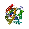
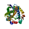
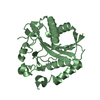
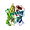
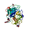
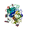
- PDB-1gky: REFINED STRUCTURE OF THE COMPLEX BETWEEN GUANYLATE KINASE AND ITS... -
+ Open data

- Basic information
| Entry | Database: PDB / ID: 1gky | |||||||||
|---|---|---|---|---|---|---|---|---|---|---|
| Title | REFINED STRUCTURE OF THE COMPLEX BETWEEN GUANYLATE KINASE AND ITS SUBSTRATE GMP AT 2.0 ANGSTROMS RESOLUTION | |||||||||
 Components Components | GUANYLATE KINASE | |||||||||
 Keywords Keywords | TRANSFERASE | |||||||||
| Function / homology |  Function and homology information Function and homology informationAzathioprine ADME / GDP biosynthetic process / guanylate kinase / Interconversion of nucleotide di- and triphosphates / purine nucleotide metabolic process / GMP kinase activity / ATP binding / nucleus / cytoplasm / cytosol Similarity search - Function | |||||||||
| Biological species |  | |||||||||
| Method |  X-RAY DIFFRACTION / Resolution: 2 Å X-RAY DIFFRACTION / Resolution: 2 Å | |||||||||
 Authors Authors | Stehle, T. / Schulz, G.E. | |||||||||
 Citation Citation | Journal: J.Mol.Biol. / Year: 1992 Title: Refined structure of the complex between guanylate kinase and its substrate GMP at 2.0 A resolution. Authors: Stehle, T. / Schulz, G.E. #1: Journal: J.Mol.Biol. / Year: 1990Title: Three-Dimensional Structure of the Complex of Guanylate Kinase from Yeast with its Substrate Gmp Authors: Stehle, T. / Schulz, G.E. #2: Journal: Eur.J.Biochem. / Year: 1989Title: Guanylate Kinase from Saccharomyces Cerevisiae. Isolation and Characterization, Crystallization and Preliminary X-Ray Analysis, Amino Acid Sequence and Comparison with Adenylate Kinases Authors: Berger, A. / Schiltz, E. / Schulz, G.E. | |||||||||
| History |
|
- Structure visualization
| Structure viewer | Molecule: MolmilJmol/JSmol |
|---|
- Downloads & links
-Download
| PDBx/mmCIF format | 1gky.cif.gz | 53.7 KB | Display | PDBx/mmCIF format |
|---|---|---|---|---|
| PDB format | pdb1gky.ent.gz | 38 KB | Display | PDB format |
| PDBx/mmJSON format | 1gky.json.gz | Tree view | PDBx/mmJSON format | |
| Others |  Other downloads Other downloads |
-Validation report
| Arichive directory | https://data.pdbj.org/pub/pdb/validation_reports/gk/1gkyftp://data.pdbj.org/pub/pdb/validation_reports/gk/1gky | HTTPS FTP |
|---|
-Related structure data
| Similar structure data |
|---|


-Links
 PDBj
PDBj

- Assembly
| Deposited unit | 
| ||||||||
|---|---|---|---|---|---|---|---|---|---|
| 1 |
| ||||||||
| Unit cell |
|
-Components
| #1: Protein | Mass: 20559.191 Da / Num. of mol.: 1 Source method: isolated from a genetically manipulated source Source: (gene. exp.) References: UniProt: P15454, guanylate kinase |
|---|---|
| #2: Chemical | ChemComp-SO4 /   Mass: 96.063 Da / Num. of mol.: 1 / Source method: obtained synthetically / Formula: SO4 Mass: 96.063 Da / Num. of mol.: 1 / Source method: obtained synthetically / Formula: SO4 |
| #3: Chemical | ChemComp-5GP /   Mass: 363.221 Da / Num. of mol.: 1 / Source method: obtained synthetically / Formula: C10H14N5O8P Mass: 363.221 Da / Num. of mol.: 1 / Source method: obtained synthetically / Formula: C10H14N5O8P |
| #4: Water | ChemComp-HOH /  Mass: 18.015 Da / Num. of mol.: 173 / Source method: isolated from a natural source / Formula: H2O Mass: 18.015 Da / Num. of mol.: 173 / Source method: isolated from a natural source / Formula: H2O |
| Has protein modification | Y |
-Experimental details
-Experiment
| Experiment | Method: X-RAY DIFFRACTION |
|---|
- Sample preparation
| Crystal | Density Matthews: 2.44 Å3/Da / Density % sol: 49.51 % | ||||||||||||||||||||||||||||||||||||||||
|---|---|---|---|---|---|---|---|---|---|---|---|---|---|---|---|---|---|---|---|---|---|---|---|---|---|---|---|---|---|---|---|---|---|---|---|---|---|---|---|---|---|
| Crystal grow | *PLUS Temperature: 20 ℃ / pH: 5.5 / Method: vapor diffusion, hanging drop | ||||||||||||||||||||||||||||||||||||||||
| Components of the solutions | *PLUS
|
-Data collection
| Radiation | Scattering type: x-ray |
|---|---|
| Radiation wavelength | Relative weight: 1 |
| Reflection | *PLUS Highest resolution: 2 Å / Num. obs: 14130 / % possible obs: 100 % / Rmerge(I) obs: 0.05 |
- Processing
| Software | Name: SIMULATED / Version: ANNEALING METHOD / Classification: refinement | ||||||||||||||||||||||||||||||||||||||||||||||||||||||||||||
|---|---|---|---|---|---|---|---|---|---|---|---|---|---|---|---|---|---|---|---|---|---|---|---|---|---|---|---|---|---|---|---|---|---|---|---|---|---|---|---|---|---|---|---|---|---|---|---|---|---|---|---|---|---|---|---|---|---|---|---|---|---|
| Refinement | Rfactor Rwork: 0.173 / Highest resolution: 2 Å | ||||||||||||||||||||||||||||||||||||||||||||||||||||||||||||
| Refinement step | Cycle: LAST / Highest resolution: 2 Å
| ||||||||||||||||||||||||||||||||||||||||||||||||||||||||||||
| Refine LS restraints |
| ||||||||||||||||||||||||||||||||||||||||||||||||||||||||||||
| Refinement | *PLUS Highest resolution: 2 Å / Lowest resolution: 7 Å / Rfactor obs: 0.173 | ||||||||||||||||||||||||||||||||||||||||||||||||||||||||||||
| Solvent computation | *PLUS | ||||||||||||||||||||||||||||||||||||||||||||||||||||||||||||
| Displacement parameters | *PLUS | ||||||||||||||||||||||||||||||||||||||||||||||||||||||||||||
| Refine LS restraints | *PLUS
|
