 Movie
Movie Controller


+ Open data

- Basic information
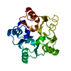
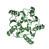
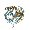
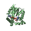
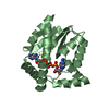
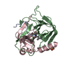
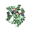
| Entry | Database: PDB / ID: 1g61 | ||||||
|---|---|---|---|---|---|---|---|
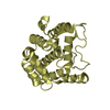
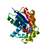
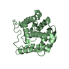
| Title | CRYSTAL STRUCTURE OF M.JANNASCHII EIF6 | ||||||
 Components Components | TRANSLATION INITIATION FACTOR 6 | ||||||
 Keywords Keywords | TRANSLATION / alpha-beta-barrel velcro closure subdomain / Structural Genomics / PSI / Protein Structure Initiative / New York SGX Research Center for Structural Genomics / NYSGXRC | ||||||
| Function / homology |  Function and homology information Function and homology informationmaturation of 5.8S rRNA / maturation of LSU-rRNA / translation initiation factor activity / cytosolic ribosome assembly / assembly of large subunit precursor of preribosome / ribosome binding / cytosol Similarity search - Function | ||||||
| Biological species |   Methanocaldococcus jannaschii (archaea) Methanocaldococcus jannaschii (archaea) | ||||||
| Method |  X-RAY DIFFRACTION / SYNCHROTRON / MAD / Resolution: 1.3 Å X-RAY DIFFRACTION / SYNCHROTRON / MAD / Resolution: 1.3 Å | ||||||
 Authors Authors | Groft, C.M. / Beckmann, R. / Sali, A. / Burley, S.K. / New York SGX Research Center for Structural Genomics (NYSGXRC) | ||||||
 Citation Citation | Journal: Nat.Struct.Biol. / Year: 2000 Title: Crystal structures of ribosome anti-association factor IF6. Authors: Groft, C.M. / Beckmann, R. / Sali, A. / Burley, S.K. | ||||||
| History |
|
- Structure visualization
| Structure viewer | Molecule: MolmilJmol/JSmol |
|---|
- Downloads & links
-Download
| PDBx/mmCIF format | 1g61.cif.gz | 122.6 KB | Display | PDBx/mmCIF format |
|---|---|---|---|---|
| PDB format | pdb1g61.ent.gz | 96.3 KB | Display | PDB format |
| PDBx/mmJSON format | 1g61.json.gz | Tree view | PDBx/mmJSON format | |
| Others |  Other downloads Other downloads |
-Validation report
| Arichive directory | https://data.pdbj.org/pub/pdb/validation_reports/g6/1g61ftp://data.pdbj.org/pub/pdb/validation_reports/g6/1g61 | HTTPS FTP |
|---|
-Related structure data
-Links
 PDBj
PDBj- Assembly
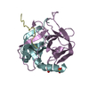
| Deposited unit | 
| ||||||||
|---|---|---|---|---|---|---|---|---|---|
| 1 | 
| ||||||||
| 2 | 
| ||||||||
| Unit cell |
|
-Components
| #1: Protein | Mass: 24481.117 Da / Num. of mol.: 2 Source method: isolated from a genetically manipulated source Source: (gene. exp.) Methanocaldococcus jannaschii (archaea)Gene: MJ0048 / Plasmid: PET28A / Species (production host): Escherichia coli / Production host:  #2: Water | ChemComp-HOH / |  Mass: 18.015 Da / Num. of mol.: 704 / Source method: isolated from a natural source / Formula: H2O Mass: 18.015 Da / Num. of mol.: 704 / Source method: isolated from a natural source / Formula: H2O |
|---|
-Experimental details
-Experiment
| Experiment | Method: X-RAY DIFFRACTION / Number of used crystals: 1 |
|---|
- Sample preparation
| Crystal | Density Matthews: 2.37 Å3/Da / Density % sol: 48.06 % | ||||||||||||||||||||||||||||||||||||||||||||||||
|---|---|---|---|---|---|---|---|---|---|---|---|---|---|---|---|---|---|---|---|---|---|---|---|---|---|---|---|---|---|---|---|---|---|---|---|---|---|---|---|---|---|---|---|---|---|---|---|---|---|
| Crystal grow | Temperature: 277 K / Method: vapor diffusion, hanging drop / pH: 5.6 Details: 31% PEG-MME 2000, 0.200M Ammonium sulfate, 0.100M MES pH 5.6, 3mM Xylitol, 10mM glycerol, VAPOR DIFFUSION, HANGING DROP, temperature 277K | ||||||||||||||||||||||||||||||||||||||||||||||||
| Crystal | *PLUS Density % sol: 48 % | ||||||||||||||||||||||||||||||||||||||||||||||||
| Crystal grow | *PLUS Temperature: 4 ℃ | ||||||||||||||||||||||||||||||||||||||||||||||||
| Components of the solutions | *PLUS
|
-Data collection
| Diffraction | Mean temperature: 193 K |
|---|---|
| Diffraction source | Source: SYNCHROTRON / Site: NSLS  / Beamline: X9B / Wavelength: 0.91825 Å / Beamline: X9B / Wavelength: 0.91825 Å |
| Detector | Type: ADSC QUANTUM 4 / Detector: CCD / Date: Feb 25, 2000 |
| Radiation | Monochromator: Silicon / Protocol: SINGLE WAVELENGTH / Monochromatic (M) / Laue (L): M / Scattering type: x-ray |
| Radiation wavelength | Wavelength: 0.91825 Å / Relative weight: 1 |
| Reflection | Resolution: 1.3→25 Å / Num. all: 110636 / Num. obs: 110636 / % possible obs: 97.7 % / Observed criterion σ(F): 0 / Observed criterion σ(I): -3 / Redundancy: 4.2 % / Biso Wilson estimate: 11 Å2 / Rmerge(I) obs: 0.036 / Rsym value: 0.036 / Net I/σ(I): 34 |
| Reflection shell | Resolution: 1.3→25 Å / Redundancy: 3.9 % / Rmerge(I) obs: 0.235 / Mean I/σ(I) obs: 6.6 / Num. unique all: 20604 / % possible all: 93 |
| Reflection | *PLUS Highest resolution: 1.3 Å / Lowest resolution: 25 Å / Num. measured all: 463351 |
| Reflection shell | *PLUS % possible obs: 95.4 % |
- Processing
| Software |
| |||||||||||||||||||||||||
|---|---|---|---|---|---|---|---|---|---|---|---|---|---|---|---|---|---|---|---|---|---|---|---|---|---|---|
| Refinement | Method to determine structure: MAD / Resolution: 1.3→25 Å / σ(F): 4 / σ(I): 0 / Stereochemistry target values: CNS defined library Details: the following residue side chains were flagged during the last round of SHELX refinement as having parameters which disagree with the restraints: 2003, 2024, 4006, 4010, 4099
| |||||||||||||||||||||||||
| Refinement step | Cycle: LAST / Resolution: 1.3→25 Å
| |||||||||||||||||||||||||
| Refine LS restraints |
| |||||||||||||||||||||||||
| LS refinement shell | Resolution: 1.3→25 Å /
| |||||||||||||||||||||||||
| Software | *PLUS Name: SHELXL-97 / Classification: refinement | |||||||||||||||||||||||||
| Refinement | *PLUS Highest resolution: 1.3 Å / Lowest resolution: 25 Å / σ(F): 4 / % reflection Rfree: 10 % | |||||||||||||||||||||||||
| Solvent computation | *PLUS | |||||||||||||||||||||||||
| Displacement parameters | *PLUS | |||||||||||||||||||||||||
| Refine LS restraints | *PLUS
|
