 Movie
Movie Controller

[English] 日本語
 Yorodumi
- PDB-2c7i: Structure of protein Ta0514, putative lipoate protein ligase from... -
+ Open data

- Basic information
| Entry | Database: PDB / ID: 2c7i | ||||||
|---|---|---|---|---|---|---|---|
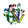
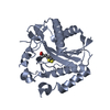
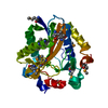
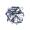
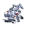
| Title | Structure of protein Ta0514, putative lipoate protein ligase from T. acidophilum. | ||||||
 Components Components | PUTATIVE LIPOATE PROTEIN LIGASE | ||||||
 Keywords Keywords | LIGASE / LIPOYLATION | ||||||
| Function / homology |  Function and homology information Function and homology informationlipoate-protein ligase / lipoate-protein ligase activity / lipoic acid binding / protein lipoylation / protein-containing complex / ATP binding / metal ion binding / cytoplasm Similarity search - Function | ||||||
| Biological species |   THERMOPLASMA ACIDOPHILUM (acidophilic) THERMOPLASMA ACIDOPHILUM (acidophilic) | ||||||
| Method |  X-RAY DIFFRACTION / SYNCHROTRON / MOLECULAR REPLACEMENT / Resolution: 2.1 Å X-RAY DIFFRACTION / SYNCHROTRON / MOLECULAR REPLACEMENT / Resolution: 2.1 Å | ||||||
 Authors Authors | Mcmanus, E. / Perham, R.N. / Luisi, B.F. | ||||||
 Citation Citation | Journal: J.Mol.Biol. / Year: 2006 Title: Structure of a Putative Lipoate Protein Ligase from Thermoplasma Acidophilum and the Mechanism of Target Selection for Post-Translational Modification. Authors: Mcmanus, E. / Luisi, B.F. / Perham, R.N. | ||||||
| History |
|
- Structure visualization
| Structure viewer | Molecule: MolmilJmol/JSmol |
|---|
- Downloads & links
-Download
| PDBx/mmCIF format | 2c7i.cif.gz | 196.8 KB | Display | PDBx/mmCIF format |
|---|---|---|---|---|
| PDB format | pdb2c7i.ent.gz | 157.5 KB | Display | PDB format |
| PDBx/mmJSON format | 2c7i.json.gz | Tree view | PDBx/mmJSON format | |
| Others |  Other downloads Other downloads |
-Validation report
| Arichive directory | https://data.pdbj.org/pub/pdb/validation_reports/c7/2c7iftp://data.pdbj.org/pub/pdb/validation_reports/c7/2c7i | HTTPS FTP |
|---|
-Related structure data


-Links
 PDBj
PDBj
- Assembly
| Deposited unit | 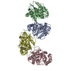
| ||||||||||||||||||||||||||||||||||||||||||||||||||||||||||||
|---|---|---|---|---|---|---|---|---|---|---|---|---|---|---|---|---|---|---|---|---|---|---|---|---|---|---|---|---|---|---|---|---|---|---|---|---|---|---|---|---|---|---|---|---|---|---|---|---|---|---|---|---|---|---|---|---|---|---|---|---|---|
| 1 | 
| ||||||||||||||||||||||||||||||||||||||||||||||||||||||||||||
| 2 | 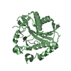
| ||||||||||||||||||||||||||||||||||||||||||||||||||||||||||||
| 3 | 
| ||||||||||||||||||||||||||||||||||||||||||||||||||||||||||||
| 4 | 
| ||||||||||||||||||||||||||||||||||||||||||||||||||||||||||||
| Unit cell |
| ||||||||||||||||||||||||||||||||||||||||||||||||||||||||||||
| Noncrystallographic symmetry (NCS) | NCS domain:
NCS domain segments: Component-ID: 1 / Beg auth comp-ID: MET / Beg label comp-ID: MET / End auth comp-ID: LEU / End label comp-ID: LEU / Auth seq-ID: 1 - 256 / Label seq-ID: 1 - 256
NCS ensembles :
|
-Components
| #1: Protein | Mass: 29915.252 Da / Num. of mol.: 4 Source method: isolated from a genetically manipulated source Source: (gene. exp.) THERMOPLASMA ACIDOPHILUM (acidophilic) / Strain: DSM 1728 / Plasmid: PET 3A / Production host:  #2: Water | ChemComp-HOH / |  Mass: 18.015 Da / Num. of mol.: 188 / Source method: isolated from a natural source / Formula: H2O Mass: 18.015 Da / Num. of mol.: 188 / Source method: isolated from a natural source / Formula: H2O |
|---|
-Experimental details
-Experiment
| Experiment | Method: X-RAY DIFFRACTION / Number of used crystals: 1 |
|---|
- Sample preparation
| Crystal | Density Matthews: 3.11 Å3/Da / Density % sol: 60.13 % Description: A STRUCTURE OF RELATIVELY POOR QUALITY DERIVED FROM A SELENOMETHIONE CONTAINING PROTEIN CRYSTAL WAS USED AS MODEL IN CONJUNCTION WITH A GOOD QUALITY NATIVE DATA SET TO GIVE THE STRUCTURE FILES DEPOSITED. |
|---|---|
| Crystal grow | pH: 6.7 / Details: pH 6.70 |
-Data collection
| Diffraction | Mean temperature: 100 K |
|---|---|
| Diffraction source | Source: SYNCHROTRON / Site: ESRF  / Beamline: ID29 / Wavelength: 1.54 / Beamline: ID29 / Wavelength: 1.54 |
| Detector | Type: ADSC CCD / Detector: CCD |
| Radiation | Protocol: SINGLE WAVELENGTH / Monochromatic (M) / Laue (L): M / Scattering type: x-ray |
| Radiation wavelength | Wavelength: 1.54 Å / Relative weight: 1 |
| Reflection | Resolution: 2.1→19.87 Å / Num. obs: 68335 / % possible obs: 95.3 % / Observed criterion σ(I): 2 / Redundancy: 10 % / Rmerge(I) obs: 0.07 / Net I/σ(I): 22 |
| Reflection shell | Resolution: 2.08→2.17 Å / Rmerge(I) obs: 0.23 / Mean I/σ(I) obs: 6 / % possible all: 72.3 |
- Processing
| Software |
| ||||||||||||||||||||||||||||||||||||||||||||||||||||||||||||||||||||||||||||||||||||||||||||||||||||||||||||||||||||||||||||||||||||||||||||||||||||||||||||||||||||||||||||||||||||||
|---|---|---|---|---|---|---|---|---|---|---|---|---|---|---|---|---|---|---|---|---|---|---|---|---|---|---|---|---|---|---|---|---|---|---|---|---|---|---|---|---|---|---|---|---|---|---|---|---|---|---|---|---|---|---|---|---|---|---|---|---|---|---|---|---|---|---|---|---|---|---|---|---|---|---|---|---|---|---|---|---|---|---|---|---|---|---|---|---|---|---|---|---|---|---|---|---|---|---|---|---|---|---|---|---|---|---|---|---|---|---|---|---|---|---|---|---|---|---|---|---|---|---|---|---|---|---|---|---|---|---|---|---|---|---|---|---|---|---|---|---|---|---|---|---|---|---|---|---|---|---|---|---|---|---|---|---|---|---|---|---|---|---|---|---|---|---|---|---|---|---|---|---|---|---|---|---|---|---|---|---|---|---|---|
| Refinement | Method to determine structure: MOLECULAR REPLACEMENT Starting model: SEE REMARK Resolution: 2.1→19.87 Å / Cor.coef. Fo:Fc: 0.95 / Cor.coef. Fo:Fc free: 0.933 / SU B: 9.866 / SU ML: 0.129 / TLS residual ADP flag: LIKELY RESIDUAL / Cross valid method: THROUGHOUT / ESU R: 0.2 / ESU R Free: 0.182 / Stereochemistry target values: MAXIMUM LIKELIHOOD Details: HYDROGENS HAVE BEEN ADDED IN THE RIDING POSITIONS. THIS ENTRY CONTAINS ATOMS WITH ZERO OCCUPANCY FOR WHICH B-FACTORS HAVE BEEN REFINED.
| ||||||||||||||||||||||||||||||||||||||||||||||||||||||||||||||||||||||||||||||||||||||||||||||||||||||||||||||||||||||||||||||||||||||||||||||||||||||||||||||||||||||||||||||||||||||
| Solvent computation | Ion probe radii: 0.8 Å / Shrinkage radii: 0.8 Å / VDW probe radii: 1.2 Å / Solvent model: MASK | ||||||||||||||||||||||||||||||||||||||||||||||||||||||||||||||||||||||||||||||||||||||||||||||||||||||||||||||||||||||||||||||||||||||||||||||||||||||||||||||||||||||||||||||||||||||
| Displacement parameters | Biso mean: 42.12 Å2
| ||||||||||||||||||||||||||||||||||||||||||||||||||||||||||||||||||||||||||||||||||||||||||||||||||||||||||||||||||||||||||||||||||||||||||||||||||||||||||||||||||||||||||||||||||||||
| Refinement step | Cycle: LAST / Resolution: 2.1→19.87 Å
| ||||||||||||||||||||||||||||||||||||||||||||||||||||||||||||||||||||||||||||||||||||||||||||||||||||||||||||||||||||||||||||||||||||||||||||||||||||||||||||||||||||||||||||||||||||||
| Refine LS restraints |
|