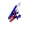
 Movie
Movie Controller


+ Open data

- Basic information
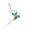
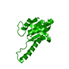
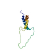
| Entry | Database: PDB / ID: 1esr | |||||||||
|---|---|---|---|---|---|---|---|---|---|---|
| Title | CRYSTAL STRUCTURE OF HUMAN MONOCYTE CHEMOTACTIC PROTEIN-2 | |||||||||
 Components Components | MONOCYTE CHEMOTACTIC PROTEIN 2 | |||||||||
 Keywords Keywords | CYTOKINE / CHEMOKINE / MONOCYTE CHEMOATTRACTANT PROTEIN / HIV-1 / PYROGLUTAMIC ACID | |||||||||
| Function / homology |  Function and homology information Function and homology informationnegative regulation of leukocyte proliferation / host-mediated suppression of viral genome replication / CCR chemokine receptor binding / eosinophil chemotaxis / chemokine activity / positive regulation of leukocyte migration / positive regulation of myoblast fusion / phospholipase activator activity / exocytosis / positive regulation of myoblast differentiation ...negative regulation of leukocyte proliferation / host-mediated suppression of viral genome replication / CCR chemokine receptor binding / eosinophil chemotaxis / chemokine activity / positive regulation of leukocyte migration / positive regulation of myoblast fusion / phospholipase activator activity / exocytosis / positive regulation of myoblast differentiation / chemokine-mediated signaling pathway / chemotaxis / response to virus / intracellular calcium ion homeostasis / calcium ion transport / antimicrobial humoral immune response mediated by antimicrobial peptide / cell-cell signaling / heparin binding / protein kinase activity / positive regulation of cell migration / inflammatory response / signal transduction / : Similarity search - Function | |||||||||
| Biological species |  Homo sapiens (human) Homo sapiens (human) | |||||||||
| Method |  X-RAY DIFFRACTION / SYNCHROTRON / MOLECULAR REPLACEMENT / Resolution: 2 Å X-RAY DIFFRACTION / SYNCHROTRON / MOLECULAR REPLACEMENT / Resolution: 2 Å | |||||||||
 Authors Authors | Blaszczyk, J. / Ji, X. | |||||||||
 Citation Citation | Journal: Biochemistry / Year: 2000 Title: Complete crystal structure of monocyte chemotactic protein-2, a CC chemokine that interacts with multiple receptors. Authors: Blaszczyk, J. / Coillie, E.V. / Proost, P. / Damme, J.V. / Opdenakker, G. / Bujacz, G.D. / Wang, J.M. / Ji, X. | |||||||||
| History |
|
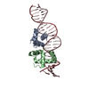
- Structure visualization
| Structure viewer | Molecule: MolmilJmol/JSmol |
|---|
- Downloads & links
-Download
| PDBx/mmCIF format | 1esr.cif.gz | 31.3 KB | Display | PDBx/mmCIF format |
|---|---|---|---|---|
| PDB format | pdb1esr.ent.gz | 19.7 KB | Display | PDB format |
| PDBx/mmJSON format | 1esr.json.gz | Tree view | PDBx/mmJSON format | |
| Others |  Other downloads Other downloads |
-Validation report
| Arichive directory | https://data.pdbj.org/pub/pdb/validation_reports/es/1esrftp://data.pdbj.org/pub/pdb/validation_reports/es/1esr | HTTPS FTP |
|---|
-Related structure data
| Related structure data |  1dokS S: Starting model for refinement |
|---|---|
| Similar structure data |


-Links
 PDBj
PDBj

- Assembly
| Deposited unit | 
| |||||||||
|---|---|---|---|---|---|---|---|---|---|---|
| 1 | 
| |||||||||
| Unit cell |
| |||||||||
| Components on special symmetry positions |
|
-Components
| #1: Protein | Mass: 8911.384 Da / Num. of mol.: 1 / Mutation: Q24(PCA) AND K69Q Source method: isolated from a genetically manipulated source Source: (gene. exp.) Homo sapiens (human) / Plasmid: PHEN1 / Production host:  |
|---|---|
| #2: Water | ChemComp-HOH /  Mass: 18.015 Da / Num. of mol.: 89 / Source method: isolated from a natural source / Formula: H2O Mass: 18.015 Da / Num. of mol.: 89 / Source method: isolated from a natural source / Formula: H2O |
| Has protein modification | Y |
-Experimental details
-Experiment
| Experiment | Method: X-RAY DIFFRACTION / Number of used crystals: 1 |
|---|
- Sample preparation
| Crystal | Density Matthews: 3.6 Å3/Da / Density % sol: 63 % | |||||||||||||||||||||||||
|---|---|---|---|---|---|---|---|---|---|---|---|---|---|---|---|---|---|---|---|---|---|---|---|---|---|---|
| Crystal grow | Temperature: 292 K / Method: vapor diffusion, hanging drop / pH: 7.5 Details: Ammonium sulfate, Tris-HCl, pH 7.5, VAPOR DIFFUSION, HANGING DROP, temperature 292K | |||||||||||||||||||||||||
| Crystal grow | *PLUS Details: drop consists of equal amounts of protein and reservoir solutions | |||||||||||||||||||||||||
| Components of the solutions | *PLUS
|
-Data collection
| Diffraction | Mean temperature: 100 K |
|---|---|
| Diffraction source | Source: SYNCHROTRON / Site: NSLS  / Beamline: X9B / Wavelength: 0.97132 / Beamline: X9B / Wavelength: 0.97132 |
| Detector | Type: ADSC QUANTUM 4 / Detector: CCD / Date: Nov 21, 1998 / Details: Mirror |
| Radiation | Monochromator: Silicon 111 / Protocol: SINGLE WAVELENGTH / Monochromatic (M) / Laue (L): M / Scattering type: x-ray |
| Radiation wavelength | Wavelength: 0.97132 Å / Relative weight: 1 |
| Reflection | Resolution: 1.9→20 Å / Num. all: 10447 / Num. obs: 10447 / % possible obs: 98.4 % / Observed criterion σ(F): 0 / Observed criterion σ(I): 0 / Redundancy: 4.1 % / Biso Wilson estimate: 32.2 Å2 / Rmerge(I) obs: 0.06 / Net I/σ(I): 19.7 |
| Reflection shell | Resolution: 1.9→1.93 Å / Redundancy: 3.6 % / Rmerge(I) obs: 0.507 / Mean I/σ(I) obs: 1.7854 / Num. unique all: 469 / % possible all: 92.7 |
| Reflection | *PLUS Num. obs: 9060 / % possible obs: 99 % / Num. measured all: 38808 / Rmerge(I) obs: 0.056 |
| Reflection shell | *PLUS % possible obs: 97.6 % / Rmerge(I) obs: 0.39 / Mean I/σ(I) obs: 3.4 |
- Processing
| Software |
| |||||||||||||||||||||||||||||||||
|---|---|---|---|---|---|---|---|---|---|---|---|---|---|---|---|---|---|---|---|---|---|---|---|---|---|---|---|---|---|---|---|---|---|---|
| Refinement | Method to determine structure: MOLECULAR REPLACEMENT Starting model: 1DOK Resolution: 2→20 Å / Num. parameters: 2793 / Num. restraintsaints: 2572 / Cross valid method: FREE R / σ(F): 0 / σ(I): 2 / Stereochemistry target values: ENGH AND HUBER Details: Least-squares refinement using the Konnert-Hendrickson conjugate-gradient algorithm
| |||||||||||||||||||||||||||||||||
| Solvent computation | Solvent model: Moews & Kretsinger, J. Mol. Biol. 91 (1975) 201-228 | |||||||||||||||||||||||||||||||||
| Refine analyze | Num. disordered residues: 1 / Occupancy sum hydrogen: 0 / Occupancy sum non hydrogen: 711 | |||||||||||||||||||||||||||||||||
| Refinement step | Cycle: LAST / Resolution: 2→20 Å
| |||||||||||||||||||||||||||||||||
| Refine LS restraints |
| |||||||||||||||||||||||||||||||||
| Software | *PLUS Name: SHELXL-97 / Classification: refinement | |||||||||||||||||||||||||||||||||
| Refinement | *PLUS | |||||||||||||||||||||||||||||||||
| Solvent computation | *PLUS | |||||||||||||||||||||||||||||||||
| Displacement parameters | *PLUS Biso mean: 49.2 Å2 |
