 Movie
Movie Controller


+ Open data

- Basic information
| Entry | Database: PDB / ID: 1eii | ||||||
|---|---|---|---|---|---|---|---|
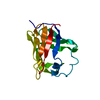
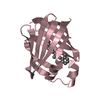
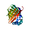
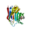
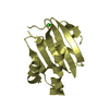
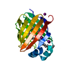
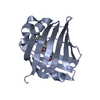
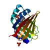
| Title | NMR STRUCTURE OF HOLO CELLULAR RETINOL-BINDING PROTEIN II | ||||||
 Components Components | CELLULAR RETINOL-BINDING PROTEIN II | ||||||
 Keywords Keywords | TRANSPORT PROTEIN / PROTEIN-LIGAND COMPLEX / BETA BARREL / HELIX-TURN-HELIX | ||||||
| Function / homology |  Function and homology information Function and homology informationRetinoid metabolism and transport / triglyceride biosynthetic process / synaptic ribbon / retinal binding / retinol binding / retinol metabolic process / molecular carrier activity / fatty acid transport / retinoid metabolic process / fatty acid binding ...Retinoid metabolism and transport / triglyceride biosynthetic process / synaptic ribbon / retinal binding / retinol binding / retinol metabolic process / molecular carrier activity / fatty acid transport / retinoid metabolic process / fatty acid binding / transmembrane transporter binding / lipid binding / nucleus / cytosol Similarity search - Function | ||||||
| Biological species |  | ||||||
| Method | SOLUTION NMR / distance geometry, simulated annealing | ||||||
 Authors Authors | Lu, J. / Lin, C.L. / Tang, C. / Ponder, J.W. / Kao, J.L. / Cistola, D.P. / Li, E. | ||||||
 Citation Citation | Journal: J.Mol.Biol. / Year: 2000 Title: Binding of retinol induces changes in rat cellular retinol-binding protein II conformation and backbone dynamics. Authors: Lu, J. / Lin, C.L. / Tang, C. / Ponder, J.W. / Kao, J.L. / Cistola, D.P. / Li, E. #1: Journal: J.Mol.Biol. / Year: 1999Title: The Structure and Dynamics of Rat Apo-Cellular Retinol-Binding Protein II in Solution: Comparison with the X-ray Structure Authors: Lu, J. / Lin, C.L. / Tang, C. / Ponder, J.W. / Kao, J.L. / Cistola, D.P. / Li, E. #2: Journal: J.Mol.Biol. / Year: 1993Title: Crystal Structures of Holo and Apo-Cellular Retinol-Binding Protein II Authors: Winter, N.S. / Bratt, J.M. / Banaszak, L.J. #3: Journal: ADV.PROTEIN CHEM. / Year: 1994Title: Lipid-Binding Proteins: A Family of Fatty Acid and Retinoid Transport Proteins Authors: Banaszak, L. / Winter, N. / Xu, Z. / Bernlohr, D.A. / Cowan, S. / Jones, T.A. #4: Journal: J.Mol.Biol. / Year: 1996Title: The NMR Solution Structure of Intestinal Fatty Acid-Binding Protein Complexed with Palmitate: Application of a Novel Distance Geometry Algorithm Authors: Hodsdon, M.E. / Ponder, J.W. / Cistola, D.P. #5: Journal: Biochemistry / Year: 1997Title: Ligand Binding Alters the Backbone Mobility of Intestinal Fatty Acid-Binding Protein as Monitored by 15N NMR Relaxation and 1H Exchange Authors: Hodsdon, M.E. / Cistola, D.P. | ||||||
| History |
|
- Structure visualization
| Structure viewer | Molecule: MolmilJmol/JSmol |
|---|
- Downloads & links
-Download
| PDBx/mmCIF format | 1eii.cif.gz | 1 MB | Display | PDBx/mmCIF format |
|---|---|---|---|---|
| PDB format | pdb1eii.ent.gz | 901.9 KB | Display | PDB format |
| PDBx/mmJSON format | 1eii.json.gz | Tree view | PDBx/mmJSON format | |
| Others |  Other downloads Other downloads |
-Validation report
| Arichive directory | https://data.pdbj.org/pub/pdb/validation_reports/ei/1eiiftp://data.pdbj.org/pub/pdb/validation_reports/ei/1eii | HTTPS FTP |
|---|
-Related structure data
| Related structure data | |
|---|---|
| Similar structure data |


-Links
 PDBj
PDBj


- Assembly
| Deposited unit | 
| |||||||||
|---|---|---|---|---|---|---|---|---|---|---|
| 1 |
| |||||||||
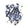
| NMR ensembles |
|
-Components
| #1: Protein | Mass: 15606.570 Da / Num. of mol.: 1 Source method: isolated from a genetically manipulated source Source: (gene. exp.)  |
|---|---|
| #2: Chemical | ChemComp-RTL /   Mass: 286.452 Da / Num. of mol.: 1 / Source method: obtained synthetically / Formula: C20H30O Mass: 286.452 Da / Num. of mol.: 1 / Source method: obtained synthetically / Formula: C20H30O |
-Experimental details
-Experiment
| Experiment | Method: SOLUTION NMR | ||||||||||||||||
|---|---|---|---|---|---|---|---|---|---|---|---|---|---|---|---|---|---|
| NMR experiment |
| ||||||||||||||||
| NMR details | Text: The structure was determined using triple-resonance NMR spectroscopy |
- Sample preparation
| Details |
| |||||||||||||||
|---|---|---|---|---|---|---|---|---|---|---|---|---|---|---|---|---|
| Sample conditions |
| |||||||||||||||
| Crystal grow | *PLUS Method: other / Details: NMR |
-NMR measurement
| NMR spectrometer |
|
|---|
- Processing
| NMR software |
| ||||||||||||
|---|---|---|---|---|---|---|---|---|---|---|---|---|---|
| Refinement | Method: distance geometry, simulated annealing / Software ordinal: 1 Details: The structure calculations were carried out using TINKER, a software package for molecular mechanics and dynamics. The protocol employs metric matrix distance geometry with pairwise Gaussian ...Details: The structure calculations were carried out using TINKER, a software package for molecular mechanics and dynamics. The protocol employs metric matrix distance geometry with pairwise Gaussian metrization followed by simulated annealing. The unique distance geometry algorithm implemented in TINKER overcomes the sampling and scaling problems of earlier distance geometry methods and is computationally more efficient. | ||||||||||||
| NMR representative | Selection criteria: closest to the average | ||||||||||||
| NMR ensemble | Conformer selection criteria: FINAL PENALTY FUNCTION VALUES WITHIN 2 STANDARD DEVIATIONS FROM THE MEAN Conformers calculated total number: 30 / Conformers submitted total number: 25 |
