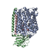
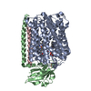
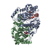
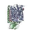
- PDB-1ehk: CRYSTAL STRUCTURE OF THE ABERRANT BA3-CYTOCHROME-C OXIDASE FROM T... -
+
Open data
ID or keywords:
Loading...
-
Basic information
Entry
Database: PDB / ID: 1ehk
Title
CRYSTAL STRUCTURE OF THE ABERRANT BA3-CYTOCHROME-C OXIDASE FROM THERMUS THERMOPHILUS
Components
(BA3-TYPE CYTOCHROME-C ...) x 3
Keywords
OXIDOREDUCTASE / cytochrome-c oxidase / membrane protein / Thermus thermophilus
Function / homology
Function and homology information
cytochrome-c oxidase / oxidative phosphorylation / cytochrome-c oxidase activity / copper ion binding / heme binding / metal ion binding / plasma membrane Similarity search - Function
Cytochrome c oxidase polypeptide 2A / Cytochrome c oxidase polypeptide 2A, ba3 type / Cytochrome c oxidase subunit IIa family / Ba3-like heme-copper oxidase subunit I / Cytochrome C oxidase subunit IIa, transmembrane domain / Cytochrome C oxidase subunit II, transmembrane / Ba3-like heme-copper oxidase subunit II, C-terminal / : / Cytochrome C Oxidase; Chain A / Cytochrome c oxidase-like, subunit I domain ...Cytochrome c oxidase polypeptide 2A / Cytochrome c oxidase polypeptide 2A, ba3 type / Cytochrome c oxidase subunit IIa family / Ba3-like heme-copper oxidase subunit I / Cytochrome C oxidase subunit IIa, transmembrane domain / Cytochrome C oxidase subunit II, transmembrane / Ba3-like heme-copper oxidase subunit II, C-terminal / : / Cytochrome C Oxidase; Chain A / Cytochrome c oxidase-like, subunit I domain / Copper centre Cu(A) / CO II and nitrous oxide reductase dinuclear copper centers signature. / Cytochrome C oxidase subunit II, transmembrane domain superfamily / Cytochrome c oxidase, subunit I, copper-binding site / Heme-copper oxidase catalytic subunit, copper B binding region signature. / Cytochrome c oxidase-like, subunit I domain / Cytochrome oxidase subunit I profile. / Cytochrome C oxidase subunit II, periplasmic domain / Cytochrome c oxidase subunit II-like C-terminal / Cytochrome oxidase subunit II copper A binding domain profile. / Cytochrome c oxidase subunit I / Cytochrome c oxidase-like, subunit I superfamily / Cytochrome C and Quinol oxidase polypeptide I / Rhopdopsin 7-helix transmembrane proteins / Rhodopsin 7-helix transmembrane proteins / Cupredoxins - blue copper proteins / Cupredoxin / Up-down Bundle / Immunoglobulin-like / Sandwich / Mainly Beta / Mainly Alpha Similarity search - Domain/homology
COPPER (II) ION / DINUCLEAR COPPER ION / HEME-AS / PROTOPORPHYRIN IX CONTAINING FE / Cytochrome c oxidase polypeptide 2A / Cytochrome c oxidase subunit 1 / Cytochrome c oxidase subunit 2 Similarity search - Component
Biological species
Thermus thermophilus (bacteria)
Method
X-RAY DIFFRACTION / SYNCHROTRON / THE STRUCTURE WAS SOLVED BY MULTIPLE ANOMALOUS DISPERSION (MAD) USING 5 DIFFERENT WAVELENGTH (2X FE-EDGE, 2X CU-EDGE, 1 REMOTE). THE DATA STATISTICS GIVEN CORRESPOND TO THE REFERENCE WAVELENGTH WHICH WAS USED FOR REFINEMENT. THE STRUCTURE WAS SOLVED WITH SHARP. / Resolution: 2.4 Å
Resolution: 2.4→2.49 Å / Rmerge(I) obs: 0.147 / Num. unique all: 7703 / % possible all: 100
Reflection
*PLUS
-
Processing
Software
Name
Version
Classification
SHARP
phasing
CNS
0.3
refinement
DENZO
datareduction
SCALEPACK
datascaling
Refinement
Method to determine structure: THE STRUCTURE WAS SOLVED BY MULTIPLE ANOMALOUS DISPERSION (MAD) USING 5 DIFFERENT WAVELENGTH (2X FE-EDGE, 2X CU-EDGE, 1 REMOTE). THE DATA STATISTICS GIVEN CORRESPOND TO ...Method to determine structure: THE STRUCTURE WAS SOLVED BY MULTIPLE ANOMALOUS DISPERSION (MAD) USING 5 DIFFERENT WAVELENGTH (2X FE-EDGE, 2X CU-EDGE, 1 REMOTE). THE DATA STATISTICS GIVEN CORRESPOND TO THE REFERENCE WAVELENGTH WHICH WAS USED FOR REFINEMENT. THE STRUCTURE WAS SOLVED WITH SHARP. Resolution: 2.4→20 Å / Rfactor Rfree error: 0.006 / Data cutoff high absF: 3262390.49 / Data cutoff high rms absF: 3262390.49 / Data cutoff low absF: 0 / Isotropic thermal model: RESTRAINED / Cross valid method: THROUGHOUT / σ(F): 0 / σ(I): 0 / Details: bulk solvent & overall anisotropic B-factors used
Rfactor
Num. reflection
% reflection
Selection details
Rfree
0.264
1972
5 %
RANDOM
Rwork
0.222
-
-
-
obs
0.222
39379
96.3 %
-
Displacement parameters
Biso mean: 50.6 Å2
Baniso -1
Baniso -2
Baniso -3
1-
9.46 Å2
0 Å2
0 Å2
2-
-
9.46 Å2
0 Å2
3-
-
-
-18.92 Å2
Refine analyze
Luzzati coordinate error obs: 0.28 Å / Luzzati d res low obs: 4 Å / Luzzati sigma a obs: 0.2 Å
Refinement step
Cycle: LAST / Resolution: 2.4→20 Å
Protein
Nucleic acid
Ligand
Solvent
Total
Num. atoms
5851
0
174
119
6144
Refine LS restraints
Refine-ID
Type
Dev ideal
Dev ideal target
X-RAY DIFFRACTION
c_bond_d
0.008
X-RAY DIFFRACTION
c_bond_d_na
X-RAY DIFFRACTION
c_bond_d_prot
X-RAY DIFFRACTION
c_angle_d
X-RAY DIFFRACTION
c_angle_d_na
X-RAY DIFFRACTION
c_angle_d_prot
X-RAY DIFFRACTION
c_angle_deg
1.6
X-RAY DIFFRACTION
c_angle_deg_na
X-RAY DIFFRACTION
c_angle_deg_prot
X-RAY DIFFRACTION
c_dihedral_angle_d
19.7
X-RAY DIFFRACTION
c_dihedral_angle_d_na
X-RAY DIFFRACTION
c_dihedral_angle_d_prot
X-RAY DIFFRACTION
c_improper_angle_d
1.09
X-RAY DIFFRACTION
c_improper_angle_d_na
X-RAY DIFFRACTION
c_improper_angle_d_prot
X-RAY DIFFRACTION
c_mcbond_it
3.1
1.5
X-RAY DIFFRACTION
c_mcangle_it
4.37
2
X-RAY DIFFRACTION
c_scbond_it
4.43
2
X-RAY DIFFRACTION
c_scangle_it
5.41
2.5
LS refinement shell
Resolution: 2.4→2.49 Å / Rfactor Rfree error: 0.021 / Total num. of bins used: 10
In the structure databanks used in Yorodumi, some data are registered as the other names, "COVID-19 virus" and "2019-nCoV". Here are the details of the virus and the list of structure data.
Jan 31, 2019. EMDB accession codes are about to change! (news from PDBe EMDB page)
EMDB accession codes are about to change! (news from PDBe EMDB page)
The allocation of 4 digits for EMDB accession codes will soon come to an end. Whilst these codes will remain in use, new EMDB accession codes will include an additional digit and will expand incrementally as the available range of codes is exhausted. The current 4-digit format prefixed with “EMD-” (i.e. EMD-XXXX) will advance to a 5-digit format (i.e. EMD-XXXXX), and so on. It is currently estimated that the 4-digit codes will be depleted around Spring 2019, at which point the 5-digit format will come into force.
The EM Navigator/Yorodumi systems omit the EMD- prefix.
Related info.:Q: What is EMD? / ID/Accession-code notation in Yorodumi/EM Navigator
Yorodumi is a browser for structure data from EMDB, PDB, SASBDB, etc.
This page is also the successor to EM Navigator detail page, and also detail information page/front-end page for Omokage search.
The word "yorodu" (or yorozu) is an old Japanese word meaning "ten thousand". "mi" (miru) is to see.
Related info.:EMDB / PDB / SASBDB / Comparison of 3 databanks / Yorodumi Search / Aug 31, 2016. New EM Navigator & Yorodumi / Yorodumi Papers / Jmol/JSmol / Function and homology information / Changes in new EM Navigator and Yorodumi
 Movie
Movie Controller

 Yorodumi
 Open data
 Basic information
 Components
 Keywords
 Function and homology information
 Thermus thermophilus (bacteria)
 X-RAY DIFFRACTION /
  Authors
 Citation
 Structure visualization
 Downloads & links
 Other downloads
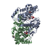


 PDBj
PDBj

 Assembly
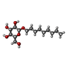
 Type: D-saccharide / Mass: 306.395 Da / Num. of mol.: 3

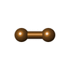

 Mass: 63.546 Da / Num. of mol.: 1 / Source method: obtained synthetically / Formula: Cu
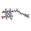
 Mass: 616.487 Da / Num. of mol.: 1 / Source method: obtained synthetically / Formula: C34H32FeN4O4
 Mass: 920.954 Da / Num. of mol.: 1 / Source method: obtained synthetically / Formula: C54H64FeN4O6
 Mass: 127.092 Da / Num. of mol.: 1 / Source method: obtained synthetically / Formula: Cu2
 Sample preparation
 / Beamline: BW6 / Wavelength: 1.05
  Processing