 Movie
Movie Controller

[English] 日本語
 Yorodumi
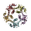
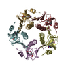
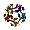
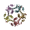
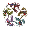
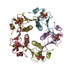
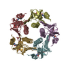
- PDB-1eei: CHOLERA TOXIN B-PENTAMER COMPLEXED WITH METANITROPHENYL-ALPHA-D-G... -
+ Open data

- Basic information
| Entry | Database: PDB / ID: 1eei | ||||||
|---|---|---|---|---|---|---|---|
| Title | CHOLERA TOXIN B-PENTAMER COMPLEXED WITH METANITROPHENYL-ALPHA-D-GALACTOSE | ||||||
 Components Components | PROTEIN (CHOLERA TOXIN B) | ||||||
 Keywords Keywords | TOXIN / ENTEROTOXIN | ||||||
| Function / homology |  Function and homology information Function and homology information | ||||||
| Biological species |   Vibrio cholerae (bacteria) Vibrio cholerae (bacteria) | ||||||
| Method |  X-RAY DIFFRACTION / MOLECULAR REPLACEMEN / Resolution: 2 Å X-RAY DIFFRACTION / MOLECULAR REPLACEMEN / Resolution: 2 Å | ||||||
 Authors Authors | Merritt, E.A. / Hol, W.G.J. | ||||||
 Citation Citation | Journal: Acta Crystallogr.,Sect.D / Year: 2001 Title: Exploration of the GM1 receptor-binding site of heat-labile enterotoxin and cholera toxin by phenyl-ring-containing galactose derivatives. Authors: Fan, E. / Merritt, E.A. / Zhang, Z. / Pickens, J.C. / Roach, C. / Ahn, M. / Hol, W.G. #1: Journal: J.Mol.Biol. / Year: 1998Title: The 1.25 A Resolution Refinement of the Cholera Toxin B-Pentamer: Evidence of Peptide Backbone Strain at the Receptor-binding Site. Authors: Merritt, E.A. / Kuhn, P. / Sarfaty, S. / Erbe, J.L. / Holmes, R.K. / Hol, W.G. #2: Journal: Structure / Year: 1997Title: Structural Foundation for the Design of Receptor Antagonists Targeting Escherichia Heat-Labile Enterotoxin Authors: Merritt, E.A. / Sarfaty, S. / Feil, I.K. / Hol, W.G.J. | ||||||
| History |
|
- Structure visualization
| Structure viewer | Molecule: MolmilJmol/JSmol |
|---|
- Downloads & links
-Download
| PDBx/mmCIF format | 1eei.cif.gz | 123.1 KB | Display | PDBx/mmCIF format |
|---|---|---|---|---|
| PDB format | pdb1eei.ent.gz | 96.5 KB | Display | PDB format |
| PDBx/mmJSON format | 1eei.json.gz | Tree view | PDBx/mmJSON format | |
| Others |  Other downloads Other downloads |
-Validation report
| Arichive directory | https://data.pdbj.org/pub/pdb/validation_reports/ee/1eeiftp://data.pdbj.org/pub/pdb/validation_reports/ee/1eei | HTTPS FTP |
|---|
-Related structure data
| Related structure data | 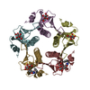 1eefC 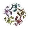 1efiC  1fd7C 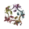 3chbS S: Starting model for refinement C: citing same article ( |
|---|---|
| Similar structure data |


-Links
 PDBj
PDBj
- Assembly
| Deposited unit | 
| ||||||||
|---|---|---|---|---|---|---|---|---|---|
| 1 |
| ||||||||
| Unit cell |
|
-Components
| #1: Protein | Mass: 11623.267 Da / Num. of mol.: 5 Source method: isolated from a genetically manipulated source Details: RECEPTOR BINDING SITE ON EACH MONOMER OCCUPIED BY METANITROPHENYL-ALPHA-D-GALACTOSIDE Source: (gene. exp.) Vibrio cholerae (bacteria) / Strain: OGAWA 41 (CLASSICAL BIOTYPE) / Production host: #2: Sugar | ChemComp-GAA / 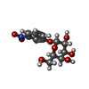  Type: D-saccharide / Mass: 301.249 Da / Num. of mol.: 5 / Source method: obtained synthetically / Formula: C12H15NO8 Type: D-saccharide / Mass: 301.249 Da / Num. of mol.: 5 / Source method: obtained synthetically / Formula: C12H15NO8#3: Water | ChemComp-HOH / |  Mass: 18.015 Da / Num. of mol.: 455 / Source method: isolated from a natural source / Formula: H2O Mass: 18.015 Da / Num. of mol.: 455 / Source method: isolated from a natural source / Formula: H2OHas protein modification | Y | |
|---|
-Experimental details
-Experiment
| Experiment | Method: X-RAY DIFFRACTION / Number of used crystals: 1 |
|---|
- Sample preparation
| Crystal | Density Matthews: 2.56 Å3/Da / Density % sol: 54.32 % | |||||||||||||||||||||||||||||||||||||||||||||
|---|---|---|---|---|---|---|---|---|---|---|---|---|---|---|---|---|---|---|---|---|---|---|---|---|---|---|---|---|---|---|---|---|---|---|---|---|---|---|---|---|---|---|---|---|---|---|
| Crystal grow | Temperature: 298 K / Method: vapor diffusion, sitting drop / pH: 4 Details: SITTING DROP, pH 4.0, VAPOR DIFFUSION, SITTING DROP, temperature 298K | |||||||||||||||||||||||||||||||||||||||||||||
| Crystal grow | *PLUS pH: 7.5 | |||||||||||||||||||||||||||||||||||||||||||||
| Components of the solutions | *PLUS
|
-Data collection
| Diffraction | Mean temperature: 100 K |
|---|---|
| Diffraction source | Source: ROTATING ANODE / Type: ENRAF-NONIUS FR591 / Wavelength: 1.5418 |
| Detector | Type: MACSCIENCE / Detector: IMAGE PLATE / Date: Apr 10, 1998 / Details: DOUBLE-FOCUS MIRRORS MIRROR |
| Radiation | Protocol: SINGLE WAVELENGTH / Monochromatic (M) / Laue (L): M / Scattering type: x-ray |
| Radiation wavelength | Wavelength: 1.5418 Å / Relative weight: 1 |
| Reflection | Resolution: 2→20 Å / Num. all: 40887 / Num. obs: 40887 / % possible obs: 93 % / Observed criterion σ(F): 0 / Observed criterion σ(I): 0 / Redundancy: 3.8 % / Rmerge(I) obs: 0.064 / Net I/σ(I): 19.2 |
| Reflection shell | Resolution: 2→2.06 Å / Redundancy: 3.5 % / Rmerge(I) obs: 0.276 / Mean I/σ(I) obs: 4.4 / Num. unique all: 3785 / % possible all: 88 |
| Reflection | *PLUS |
| Reflection shell | *PLUS % possible obs: 88 % |
- Processing
| Software |
| |||||||||||||||||||||||||
|---|---|---|---|---|---|---|---|---|---|---|---|---|---|---|---|---|---|---|---|---|---|---|---|---|---|---|
| Refinement | Method to determine structure: MOLECULAR REPLACEMEN Starting model: PDB ENTRY 3CHB Resolution: 2→20 Å / σ(F): 1 / σ(I): 0 / Stereochemistry target values: ENGH & HUBER
| |||||||||||||||||||||||||
| Refinement step | Cycle: LAST / Resolution: 2→20 Å
| |||||||||||||||||||||||||
| Refine LS restraints |
| |||||||||||||||||||||||||
| Software | *PLUS Name: X-PLOR / Version: 3.851 / Classification: refinement | |||||||||||||||||||||||||
| Refinement | *PLUS Highest resolution: 2 Å / Lowest resolution: 20 Å / σ(F): 1 | |||||||||||||||||||||||||
| Solvent computation | *PLUS | |||||||||||||||||||||||||
| Displacement parameters | *PLUS |
