 Movie
Movie Controller

[English] 日本語
 Yorodumi
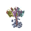
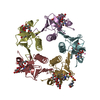
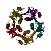
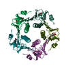
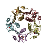
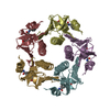
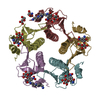
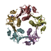
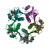
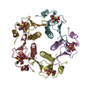
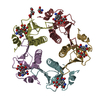
- PDB-1djr: HEAT-LABILE ENTEROTOXIN B-PENTAMER COMPLEXED WITH M-CARBOXYPHENYL... -
+ Open data

- Basic information
| Entry | Database: PDB / ID: 1djr | ||||||
|---|---|---|---|---|---|---|---|
| Title | HEAT-LABILE ENTEROTOXIN B-PENTAMER COMPLEXED WITH M-CARBOXYPHENYL-ALPHA-D-GALACTOSE | ||||||
 Components Components | HEAT-LABILE ENTEROTOXIN | ||||||
 Keywords Keywords | TOXIN / AB5 TOXINS / CELL RECOGNITION / SIX-STRANDED ANTIPARALLEL BETA-SHEET | ||||||
| Function / homology |  Function and homology information Function and homology informationSecretion of toxins / toxin activity / killing of cells of another organism / extracellular region Similarity search - Function | ||||||
| Biological species |  | ||||||
| Method |  X-RAY DIFFRACTION / SYNCHROTRON / MOLECULAR REPLACEMENT / Resolution: 1.3 Å X-RAY DIFFRACTION / SYNCHROTRON / MOLECULAR REPLACEMENT / Resolution: 1.3 Å | ||||||
 Authors Authors | Minke, W.E. / Pickens, J. / Merritt, E.A. / Fan, E. / Verlinde, C.L.M.J. / Hol, W.G.J. | ||||||
 Citation Citation | Journal: Acta Crystallogr.,Sect.D / Year: 2000 Title: Structure of m-carboxyphenyl-alpha-D-galactopyranoside complexed to heat-labile enterotoxin at 1.3 A resolution: surprising variations in ligand-binding modes. Authors: Minke, W.E. / Pickens, J. / Merritt, E.A. / Fan, E. / Verlinde, C.L. / Hol, W.G. | ||||||
| History |
|
- Structure visualization
| Structure viewer | Molecule: MolmilJmol/JSmol |
|---|
- Downloads & links
-Download
| PDBx/mmCIF format | 1djr.cif.gz | 254.6 KB | Display | PDBx/mmCIF format |
|---|---|---|---|---|
| PDB format | pdb1djr.ent.gz | 204.5 KB | Display | PDB format |
| PDBx/mmJSON format | 1djr.json.gz | Tree view | PDBx/mmJSON format | |
| Others |  Other downloads Other downloads |
-Validation report
| Arichive directory | https://data.pdbj.org/pub/pdb/validation_reports/dj/1djrftp://data.pdbj.org/pub/pdb/validation_reports/dj/1djr | HTTPS FTP |
|---|
-Related structure data
| Related structure data |  1lt5S S: Starting model for refinement |
|---|---|
| Similar structure data |


-Links
 PDBj
PDBj
- Assembly
| Deposited unit | 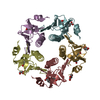
| ||||||||
|---|---|---|---|---|---|---|---|---|---|
| 1 |
| ||||||||
| Unit cell |
|
-Components
| #1: Protein | Mass: 11807.539 Da / Num. of mol.: 5 / Fragment: B PENTAMER Source method: isolated from a genetically manipulated source Source: (gene. exp.) #2: Sugar | ChemComp-GLA / 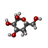  Type: D-saccharide, alpha linking / Mass: 180.156 Da / Num. of mol.: 5 / Source method: obtained synthetically / Formula: C6H12O6 Type: D-saccharide, alpha linking / Mass: 180.156 Da / Num. of mol.: 5 / Source method: obtained synthetically / Formula: C6H12O6#3: Chemical |   Mass: 92.094 Da / Num. of mol.: 3 / Source method: obtained synthetically / Formula: C3H8O3 Mass: 92.094 Da / Num. of mol.: 3 / Source method: obtained synthetically / Formula: C3H8O3#4: Chemical |   Mass: 122.121 Da / Num. of mol.: 3 / Source method: obtained synthetically / Formula: C7H6O2 Mass: 122.121 Da / Num. of mol.: 3 / Source method: obtained synthetically / Formula: C7H6O2#5: Water | ChemComp-HOH / |  Mass: 18.015 Da / Num. of mol.: 807 / Source method: isolated from a natural source / Formula: H2O Mass: 18.015 Da / Num. of mol.: 807 / Source method: isolated from a natural source / Formula: H2OCompound details | different binding modes for the ligand in the five binding sites | Has protein modification | Y | |
|---|
-Experimental details
-Experiment
| Experiment | Method: X-RAY DIFFRACTION / Number of used crystals: 1 |
|---|
- Sample preparation
| Crystal | Density Matthews: 2.2 Å3/Da / Density % sol: 46 % | ||||||||||||||||||||||||||||||||||||||||||||||||||||||||||||
|---|---|---|---|---|---|---|---|---|---|---|---|---|---|---|---|---|---|---|---|---|---|---|---|---|---|---|---|---|---|---|---|---|---|---|---|---|---|---|---|---|---|---|---|---|---|---|---|---|---|---|---|---|---|---|---|---|---|---|---|---|---|
| Crystal grow | Temperature: 298 K / Method: liquid diffusion / pH: 7.5 Details: PEG 6000, NaCl, Tris-HCl, pH 7.5, LIQUID DIFFUSION, temperature 298K | ||||||||||||||||||||||||||||||||||||||||||||||||||||||||||||
| Crystal grow | *PLUS Method: three-layer capillary method | ||||||||||||||||||||||||||||||||||||||||||||||||||||||||||||
| Components of the solutions | *PLUS
|
-Data collection
| Diffraction | Mean temperature: 100 K |
|---|---|
| Diffraction source | Source: SYNCHROTRON / Site: SSRL  / Beamline: BL9-1 / Wavelength: 0.98 / Beamline: BL9-1 / Wavelength: 0.98 |
| Detector | Type: MARRESEARCH / Detector: IMAGE PLATE / Date: Jun 16, 1999 / Details: mirror |
| Radiation | Monochromator: SI311 / Protocol: SINGLE WAVELENGTH / Monochromatic (M) / Laue (L): M / Scattering type: x-ray |
| Radiation wavelength | Wavelength: 0.98 Å / Relative weight: 1 |
| Reflection | Resolution: 1.3→20 Å / Num. all: 127782 / Num. obs: 127782 / % possible obs: 99.4 % / Redundancy: 2.9 % / Biso Wilson estimate: 9.382 Å2 / Rmerge(I) obs: 0.046 / Net I/σ(I): 14.9 |
| Reflection shell | Resolution: 1.3→1.35 Å / Redundancy: 2.3 % / Rmerge(I) obs: 0.465 / Mean I/σ(I) obs: 2.2 / Num. unique all: 12229 / % possible all: 95.5 |
| Reflection | *PLUS Num. measured all: 837038 |
| Reflection shell | *PLUS % possible obs: 95.5 % |
- Processing
| Software |
| |||||||||||||||||||||||||||||||||
|---|---|---|---|---|---|---|---|---|---|---|---|---|---|---|---|---|---|---|---|---|---|---|---|---|---|---|---|---|---|---|---|---|---|---|
| Refinement | Method to determine structure: MOLECULAR REPLACEMENT Starting model: 1LT5 Resolution: 1.3→20 Å / Num. parameters: 45729 / Num. restraintsaints: 54312 / Cross valid method: FREE R / Stereochemistry target values: ENGH AND HUBER
| |||||||||||||||||||||||||||||||||
| Solvent computation | Solvent model: MOEWS & KRETSINGER, J.MOL.BIOL.91(1975)201-228 | |||||||||||||||||||||||||||||||||
| Refine analyze | Num. disordered residues: 20 / Occupancy sum hydrogen: 4171 / Occupancy sum non hydrogen: 5018 | |||||||||||||||||||||||||||||||||
| Refinement step | Cycle: LAST / Resolution: 1.3→20 Å
| |||||||||||||||||||||||||||||||||
| Refine LS restraints |
|
