 Movie
Movie Controller


+ Open data

- Basic information
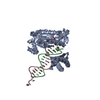
| Entry | Database: PDB / ID: 1ecy | ||||||||||||
|---|---|---|---|---|---|---|---|---|---|---|---|---|---|
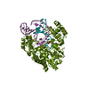
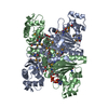
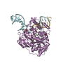
| Title | PROTEASE INHIBITOR ECOTIN | ||||||||||||
 Components Components | ECOTIN | ||||||||||||
 Keywords Keywords | PROTEASE INHIBITOR / BETA-SHEET STRUCTURE / SERINE PROTEASE INHIBITOR / PERIPLASMIC | ||||||||||||
| Function / homology |  Function and homology information Function and homology informationserine-type endopeptidase inhibitor activity / defense response / outer membrane-bounded periplasmic space / protein homodimerization activity Similarity search - Function | ||||||||||||
| Biological species |  | ||||||||||||
| Method |  X-RAY DIFFRACTION / SYNCHROTRON / MOLECULAR REPLACEMENT / Resolution: 2.19 Å X-RAY DIFFRACTION / SYNCHROTRON / MOLECULAR REPLACEMENT / Resolution: 2.19 Å | ||||||||||||
 Authors Authors | Shin, D.H. / Suh, S.W. | ||||||||||||
 Citation Citation | Journal: Protein Sci. / Year: 1996 Title: Crystal structure analyses of uncomplexed ecotin in two crystal forms: implications for its function and stability. Authors: Shin, D.H. / Song, H.K. / Seong, I.S. / Lee, C.S. / Chung, C.H. / Suh, S.W. #1: Journal: J.Mol.Biol. / Year: 1993Title: Crystallization and Preliminary X-Ray Crystallographic Analysis of the Protease Inhibitor Ecotin Authors: Shin, D.H. / Hwang, K.Y. / Kim, K.K. / Lee, H.R. / Lee, C.S. / Chung, C.H. / Suh, S.W. | ||||||||||||
| History |
|
- Structure visualization
| Structure viewer | Molecule: MolmilJmol/JSmol |
|---|
- Downloads & links
-Download
| PDBx/mmCIF format | 1ecy.cif.gz | 52.4 KB | Display | PDBx/mmCIF format |
|---|---|---|---|---|
| PDB format | pdb1ecy.ent.gz | 39.2 KB | Display | PDB format |
| PDBx/mmJSON format | 1ecy.json.gz | Tree view | PDBx/mmJSON format | |
| Others |  Other downloads Other downloads |
-Validation report
| Arichive directory | https://data.pdbj.org/pub/pdb/validation_reports/ec/1ecyftp://data.pdbj.org/pub/pdb/validation_reports/ec/1ecy | HTTPS FTP |
|---|
-Related structure data


-Links
 PDBj
PDBj


- Assembly
| Deposited unit | 
| ||||||||
|---|---|---|---|---|---|---|---|---|---|
| 1 | 
| ||||||||
| Unit cell |
|
-Components
-Protein / Non-polymers , 2 types, 81 molecules A
| #1: Protein | Mass: 16120.507 Da / Num. of mol.: 1 Source method: isolated from a genetically manipulated source Details: ESCHERICHIA COLI PROTEASE INHIBITOR / Source: (gene. exp.) |
|---|---|
| #6: Water | ChemComp-HOH / Mass: 18.015 Da / Num. of mol.: 80 / Source method: isolated from a natural source / Formula: H2O |
-Sugars , 4 types, 18 molecules 

| #2: Polysaccharide | alpha-D-glucopyranose-(1-1)-alpha-D-glucopyranose 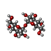  Source method: isolated from a genetically manipulated source Details: oligosaccharide with reducing-end-to-reducing-end glycosidic bond References: trehalose #3: Polysaccharide | alpha-D-glucopyranose-(1-1)-alpha-D-glucopyranose Source method: isolated from a genetically manipulated source Details: oligosaccharide with reducing-end-to-reducing-end glycosidic bond References: trehalose #4: Sugar | ChemComp-GLC /  Type: D-saccharide, alpha linking / Mass: 180.156 Da / Num. of mol.: 4 Type: D-saccharide, alpha linking / Mass: 180.156 Da / Num. of mol.: 4Source method: isolated from a genetically manipulated source Formula: C6H12O6 #5: Sugar | ChemComp-BGC / |  Type: D-saccharide, beta linking / Mass: 180.156 Da / Num. of mol.: 1 Type: D-saccharide, beta linking / Mass: 180.156 Da / Num. of mol.: 1Source method: isolated from a genetically manipulated source Formula: C6H12O6 |
|---|
-Details
| Has protein modification | Y |
|---|
-Experimental details
-Experiment
| Experiment | Method: X-RAY DIFFRACTION / Number of used crystals: 1 |
|---|
- Sample preparation
| Crystal | Density Matthews: 2.62 Å3/Da / Density % sol: 53.1 % | ||||||||||||||||||||||||||||||||||||||||
|---|---|---|---|---|---|---|---|---|---|---|---|---|---|---|---|---|---|---|---|---|---|---|---|---|---|---|---|---|---|---|---|---|---|---|---|---|---|---|---|---|---|
| Crystal grow | *PLUS Temperature: 22.5-23.5 ℃ / pH: 8.5 / Method: vapor diffusion, hanging drop | ||||||||||||||||||||||||||||||||||||||||
| Components of the solutions | *PLUS
|
-Data collection
| Diffraction | Mean temperature: 287 K |
|---|---|
| Diffraction source | Source: SYNCHROTRON / Site: Photon Factory  / Beamline: BL-6A / Wavelength: 1 / Beamline: BL-6A / Wavelength: 1 |
| Detector | Type: FUJI / Detector: IMAGE PLATE / Date: Jun 24, 1994 |
| Radiation | Monochromatic (M) / Laue (L): M / Scattering type: x-ray |
| Radiation wavelength | Wavelength: 1 Å / Relative weight: 1 |
| Reflection | Resolution: 2.19→84 Å / Num. obs: 8537 / % possible obs: 91.6 % / Observed criterion σ(I): 3 / Redundancy: 7.9 % / Rmerge(I) obs: 0.073 |
| Reflection shell | Resolution: 2.19→2.29 Å / % possible all: 73.2 |
| Reflection shell | *PLUS % possible obs: 73.2 % |
- Processing
| Software |
| ||||||||||||||||||||||||||||||||||||||||||||||||||||||||||||
|---|---|---|---|---|---|---|---|---|---|---|---|---|---|---|---|---|---|---|---|---|---|---|---|---|---|---|---|---|---|---|---|---|---|---|---|---|---|---|---|---|---|---|---|---|---|---|---|---|---|---|---|---|---|---|---|---|---|---|---|---|---|
| Refinement | Method to determine structure: MOLECULAR REPLACEMENT Starting model: ECOTIN PART OF ECOTIN-TRYPSIN COMPLEX Resolution: 2.19→6 Å / σ(F): 2
| ||||||||||||||||||||||||||||||||||||||||||||||||||||||||||||
| Displacement parameters | Biso mean: 36 Å2 | ||||||||||||||||||||||||||||||||||||||||||||||||||||||||||||
| Refinement step | Cycle: LAST / Resolution: 2.19→6 Å
| ||||||||||||||||||||||||||||||||||||||||||||||||||||||||||||
| Refine LS restraints |
| ||||||||||||||||||||||||||||||||||||||||||||||||||||||||||||
| Software | *PLUS Name: X-PLOR / Version: 3.1 / Classification: refinement | ||||||||||||||||||||||||||||||||||||||||||||||||||||||||||||
| Refine LS restraints | *PLUS
|
