 Movie
Movie Controller

[English] 日本語
 Yorodumi
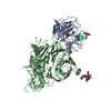
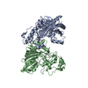
- PDB-1ecb: ESCHERICHIA COLI GLUTAMINE PHOSPHORIBOSYLPYROPHOSPHATE (PRPP) AMI... -
+ Open data

- Basic information
| Entry | Database: PDB / ID: 1ecb | |||||||||
|---|---|---|---|---|---|---|---|---|---|---|
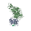
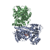
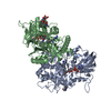
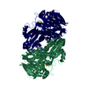
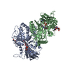
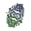
| Title | ESCHERICHIA COLI GLUTAMINE PHOSPHORIBOSYLPYROPHOSPHATE (PRPP) AMIDOTRANSFERASE COMPLEXED WITH 2 GMP, 1 MG PER SUBUNIT | |||||||||
 Components Components | GLUTAMINE PHOSPHORIBOSYLPYROPHOSPHATE AMIDOTRANSFERASE | |||||||||
 Keywords Keywords | TRANSFERASE / GLUTAMINE AMIDOTRANSFERASE / PURINE BIOSYNTHESIS / GLYCOSYLTRANSFERASE / GMP / GUANINE 5'-MONOPHOSPHATE | |||||||||
| Function / homology |  Function and homology information Function and homology informationamidophosphoribosyltransferase / amidophosphoribosyltransferase activity / purine nucleobase biosynthetic process / L-glutamine metabolic process / purine nucleotide biosynthetic process / 'de novo' IMP biosynthetic process / guanosine tetraphosphate binding / glycosyltransferase activity / magnesium ion binding / identical protein binding ...amidophosphoribosyltransferase / amidophosphoribosyltransferase activity / purine nucleobase biosynthetic process / L-glutamine metabolic process / purine nucleotide biosynthetic process / 'de novo' IMP biosynthetic process / guanosine tetraphosphate binding / glycosyltransferase activity / magnesium ion binding / identical protein binding / cytosol / cytoplasm Similarity search - Function | |||||||||
| Biological species |  | |||||||||
| Method |  X-RAY DIFFRACTION / MOLECULAR REPLACEMENT / Resolution: 2.7 Å X-RAY DIFFRACTION / MOLECULAR REPLACEMENT / Resolution: 2.7 Å | |||||||||
 Authors Authors | Krahn, J.M. / Smith, J.L. | |||||||||
 Citation Citation | Journal: Biochemistry / Year: 1997 Title: Coupled formation of an amidotransferase interdomain ammonia channel and a phosphoribosyltransferase active site. Authors: Krahn, J.M. / Kim, J.H. / Burns, M.R. / Parry, R.J. / Zalkin, H. / Smith, J.L. | |||||||||
| History |
|
- Structure visualization
| Structure viewer | Molecule: MolmilJmol/JSmol |
|---|
- Downloads & links
-Download
| PDBx/mmCIF format | 1ecb.cif.gz | 378 KB | Display | PDBx/mmCIF format |
|---|---|---|---|---|
| PDB format | pdb1ecb.ent.gz | 308.3 KB | Display | PDB format |
| PDBx/mmJSON format | 1ecb.json.gz | Tree view | PDBx/mmJSON format | |
| Others |  Other downloads Other downloads |
-Validation report
| Arichive directory | https://data.pdbj.org/pub/pdb/validation_reports/ec/1ecbftp://data.pdbj.org/pub/pdb/validation_reports/ec/1ecb | HTTPS FTP |
|---|
-Related structure data
| Related structure data | 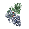 1eccC 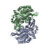 1ecfS S: Starting model for refinement C: citing same article ( |
|---|---|
| Similar structure data |


-Links
 PDBj
PDBj


- Assembly
| Deposited unit | 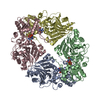
| ||||||||||||||||
|---|---|---|---|---|---|---|---|---|---|---|---|---|---|---|---|---|---|
| 1 | 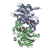
| ||||||||||||||||
| 2 | 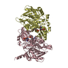
| ||||||||||||||||
| 3 |
| ||||||||||||||||
| 4 | 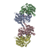
| ||||||||||||||||
| Unit cell |
| ||||||||||||||||
| Noncrystallographic symmetry (NCS) | NCS oper:
|
-Components
| #1: Protein | Mass: 56422.684 Da / Num. of mol.: 4 Source method: isolated from a genetically manipulated source Source: (gene. exp.) References: UniProt: P00496, UniProt: P0AG16*PLUS, amidophosphoribosyltransferase #2: Chemical | ChemComp-MG /   Mass: 24.305 Da / Num. of mol.: 4 / Source method: obtained synthetically / Formula: Mg Mass: 24.305 Da / Num. of mol.: 4 / Source method: obtained synthetically / Formula: Mg#3: Chemical | ChemComp-5GP /   Mass: 363.221 Da / Num. of mol.: 8 / Source method: obtained synthetically / Formula: C10H14N5O8P Mass: 363.221 Da / Num. of mol.: 8 / Source method: obtained synthetically / Formula: C10H14N5O8P#4: Water | ChemComp-HOH / |  Mass: 18.015 Da / Num. of mol.: 39 / Source method: isolated from a natural source / Formula: H2O Mass: 18.015 Da / Num. of mol.: 39 / Source method: isolated from a natural source / Formula: H2OCompound details | THE GUANINE BASE OF GMP BOUND IN THE PRTASE ACTIVE SITE IS NOT WELL ORDERED, AND APPEARS TO WOBBLE ...THE GUANINE BASE OF GMP BOUND IN THE PRTASE ACTIVE SITE IS NOT WELL ORDERED, AND APPEARS TO WOBBLE IN THE PLANE OF THE BASE. ONLY A SINGLE CONFORMER HAS BEEN INCLUDED IN THIS MODEL TO THE LIMITED RESOLUTION | Has protein modification | N | |
|---|
-Experimental details
-Experiment
| Experiment | Method: X-RAY DIFFRACTION / Number of used crystals: 1 |
|---|
- Sample preparation
| Crystal | Density Matthews: 2.63 Å3/Da / Density % sol: 53 % | ||||||||||||||||||||||||||||||||||||||||||||||||||||||||||||||||||||||||||||||
|---|---|---|---|---|---|---|---|---|---|---|---|---|---|---|---|---|---|---|---|---|---|---|---|---|---|---|---|---|---|---|---|---|---|---|---|---|---|---|---|---|---|---|---|---|---|---|---|---|---|---|---|---|---|---|---|---|---|---|---|---|---|---|---|---|---|---|---|---|---|---|---|---|---|---|---|---|---|---|---|
| Crystal grow | pH: 6 Details: 5 MM GMP, 20 MM MGCL2, 100 MM BIS-TRIS PH 6.0, 10% PEG-3350, 10% 2-PROPANOL | ||||||||||||||||||||||||||||||||||||||||||||||||||||||||||||||||||||||||||||||
| Crystal | *PLUS | ||||||||||||||||||||||||||||||||||||||||||||||||||||||||||||||||||||||||||||||
| Crystal grow | *PLUS Method: vapor diffusion | ||||||||||||||||||||||||||||||||||||||||||||||||||||||||||||||||||||||||||||||
| Components of the solutions | *PLUS
|
-Data collection
| Diffraction | Mean temperature: 110 K |
|---|---|
| Diffraction source | Source: ROTATING ANODE / Type: RIGAKU RUH2R / Wavelength: 1.5418 |
| Detector | Type: RIGAKU / Detector: IMAGE PLATE / Date: Mar 1, 1996 |
| Radiation | Monochromator: MSC DOUBLE MIRROR / Monochromatic (M) / Laue (L): M / Scattering type: x-ray |
| Radiation wavelength | Wavelength: 1.5418 Å / Relative weight: 1 |
| Reflection | Resolution: 2.7→30 Å / Num. obs: 58955 / % possible obs: 97.7 % / Observed criterion σ(I): -3 / Redundancy: 6 % / Rsym value: 0.092 / Net I/σ(I): 12.7 |
| Reflection shell | Resolution: 2.7→2.8 Å / Mean I/σ(I) obs: 3.5 / Rsym value: 0.33 / % possible all: 78.9 |
| Reflection | *PLUS Num. measured all: 354923 / Rmerge(I) obs: 0.092 |
| Reflection shell | *PLUS % possible obs: 78.9 % / Rmerge(I) obs: 0.33 |
- Processing
| Software |
| ||||||||||||||||||||||||||||||||||||||||||||||||||||||||||||||||||||||||||||||||
|---|---|---|---|---|---|---|---|---|---|---|---|---|---|---|---|---|---|---|---|---|---|---|---|---|---|---|---|---|---|---|---|---|---|---|---|---|---|---|---|---|---|---|---|---|---|---|---|---|---|---|---|---|---|---|---|---|---|---|---|---|---|---|---|---|---|---|---|---|---|---|---|---|---|---|---|---|---|---|---|---|---|
| Refinement | Method to determine structure: MOLECULAR REPLACEMENT Starting model: PDB ENTRY 1ECF Resolution: 2.7→15 Å / Data cutoff high absF: 1000000 / Data cutoff low absF: 0 / Cross valid method: FREE R, THROUGHOUT / σ(F): 0
| ||||||||||||||||||||||||||||||||||||||||||||||||||||||||||||||||||||||||||||||||
| Displacement parameters | Biso mean: 32 Å2 | ||||||||||||||||||||||||||||||||||||||||||||||||||||||||||||||||||||||||||||||||
| Refinement step | Cycle: LAST / Resolution: 2.7→15 Å
| ||||||||||||||||||||||||||||||||||||||||||||||||||||||||||||||||||||||||||||||||
| Refine LS restraints |
| ||||||||||||||||||||||||||||||||||||||||||||||||||||||||||||||||||||||||||||||||
| Refine LS restraints NCS | NCS model details: RESTRAINTS BETWEEN SUBUNIT FRAGMENTS | ||||||||||||||||||||||||||||||||||||||||||||||||||||||||||||||||||||||||||||||||
| LS refinement shell | Resolution: 2.7→2.8 Å / Total num. of bins used: 10
| ||||||||||||||||||||||||||||||||||||||||||||||||||||||||||||||||||||||||||||||||
| Xplor file |
| ||||||||||||||||||||||||||||||||||||||||||||||||||||||||||||||||||||||||||||||||
| Software | *PLUS Name: X-PLOR / Version: 3.8 / Classification: refinement | ||||||||||||||||||||||||||||||||||||||||||||||||||||||||||||||||||||||||||||||||
| Refine LS restraints | *PLUS
|
