 Movie
Movie Controller

[English] 日本語
 Yorodumi
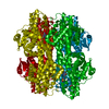
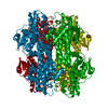
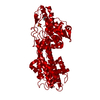
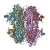
- PDB-1eb4: Histidine Ammonia-Lyase (HAL) Mutant F329A from Pseudomonas putida -
+ Open data

- Basic information
| Entry | Database: PDB / ID: 1eb4 | |||||||||
|---|---|---|---|---|---|---|---|---|---|---|
| Title | Histidine Ammonia-Lyase (HAL) Mutant F329A from Pseudomonas putida | |||||||||
 Components Components | HISTIDINE AMMONIA-LYASE | |||||||||
 Keywords Keywords | LYASE / AMMONIA-LYASE / HISTIDINE DEGRADATION | |||||||||
| Function / homology |  Function and homology information Function and homology informationhistidine ammonia-lyase / histidine ammonia-lyase activity / L-histidine catabolic process to glutamate and formamide / L-histidine catabolic process to glutamate and formate / cytoplasm Similarity search - Function | |||||||||
| Biological species |  PSEUDOMONAS PUTIDA (bacteria) PSEUDOMONAS PUTIDA (bacteria) | |||||||||
| Method |  X-RAY DIFFRACTION / OTHER / Resolution: 2 Å X-RAY DIFFRACTION / OTHER / Resolution: 2 Å | |||||||||
 Authors Authors | Baedeker, M. / Schulz, G.E. | |||||||||
 Citation Citation | Journal: Structure / Year: 2002 Title: Autocatalytic Peptide Cyclization During Chain Folding of Histidine Ammonia-Lyase. Authors: Baedeker, M. / Schulz, G.E. #1: Journal: Biochemistry / Year: 1999Title: Crystal Structure of Histidine Ammonia-Lyase Revealing a Novel Polypeptide Modification as the Catalytic Electrophile Authors: Schwede, T.F. / Retey, J. / Schulz, G.E. #2: Journal: Protein Eng. / Year: 1999 Title: Homogenization and Crystallization of Histidine Ammonia-Lyase by Exchange of a Surface Cysteine Residue Authors: Schwede, T.F. / Baedeker, M. / Langer, M. / Retey, J. / Schulz, G.E. | |||||||||
| History |
|
- Structure visualization
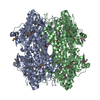
| Structure viewer | Molecule: MolmilJmol/JSmol |
|---|
- Downloads & links
-Download
| PDBx/mmCIF format | 1eb4.cif.gz | 115.8 KB | Display | PDBx/mmCIF format |
|---|---|---|---|---|
| PDB format | pdb1eb4.ent.gz | 88 KB | Display | PDB format |
| PDBx/mmJSON format | 1eb4.json.gz | Tree view | PDBx/mmJSON format | |
| Others |  Other downloads Other downloads |
-Validation report
| Summary document | 1eb4_validation.pdf.gz | 452.9 KB | Display | wwPDB validaton report |
|---|---|---|---|---|
| Full document | 1eb4_full_validation.pdf.gz | 462 KB | Display | |
| Data in XML | 1eb4_validation.xml.gz | 24.4 KB | Display | |
| Data in CIF | 1eb4_validation.cif.gz | 36 KB | Display | |
| Arichive directory | https://data.pdbj.org/pub/pdb/validation_reports/eb/1eb4ftp://data.pdbj.org/pub/pdb/validation_reports/eb/1eb4 | HTTPS FTP |
-Related structure data


-Links
 PDBj
PDBj

- Assembly
| Deposited unit | 
| ||||||||||||
|---|---|---|---|---|---|---|---|---|---|---|---|---|---|
| 1 | 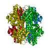
| ||||||||||||
| Unit cell |
| ||||||||||||
| Components on special symmetry positions |
|
-Components
| #1: Protein | Mass: 53543.129 Da / Num. of mol.: 1 / Mutation: YES Source method: isolated from a genetically manipulated source Details: ALA 142, SER 143 AND GLY 144 ARE FORMING AN 4-METHYLIDENE-IMIDAZOLE-5-ONE GROUP (MDO).THE CARBONYL CARBON OF ALA 142 IS BONDED TO THE NITROGEN OF GLY 144. THE CARBONYL OXYGEN OF ALA 142 IS ...Details: ALA 142, SER 143 AND GLY 144 ARE FORMING AN 4-METHYLIDENE-IMIDAZOLE-5-ONE GROUP (MDO).THE CARBONYL CARBON OF ALA 142 IS BONDED TO THE NITROGEN OF GLY 144. THE CARBONYL OXYGEN OF ALA 142 IS DELETED. THE SIDE CHAIN OF SER 143 IS DEHYDRATED. Source: (gene. exp.) PSEUDOMONAS PUTIDA (bacteria) / Cellular location: CYTOPLASM / Gene: HUTH / Plasmid: PT7-7H / Production host: | ||
|---|---|---|---|
| #2: Chemical | ChemComp-SO4 /   Mass: 96.063 Da / Num. of mol.: 1 / Source method: obtained synthetically / Formula: SO4 Mass: 96.063 Da / Num. of mol.: 1 / Source method: obtained synthetically / Formula: SO4 | ||
| #3: Chemical | ChemComp-GOL /   Mass: 92.094 Da / Num. of mol.: 1 / Source method: obtained synthetically / Formula: C3H8O3 Mass: 92.094 Da / Num. of mol.: 1 / Source method: obtained synthetically / Formula: C3H8O3 | ||
| #4: Water | ChemComp-HOH /  Mass: 18.015 Da / Num. of mol.: 387 / Source method: isolated from a natural source / Formula: H2O Mass: 18.015 Da / Num. of mol.: 387 / Source method: isolated from a natural source / Formula: H2O | ||
| Compound details | ENGINEERED| Has protein modification | Y | |
-Experimental details
-Experiment
| Experiment | Method: X-RAY DIFFRACTION / Number of used crystals: 1 |
|---|
- Sample preparation
| Crystal | Density Matthews: 2.78 Å3/Da / Density % sol: 55.83 % | ||||||||||||||||||||||||||||||||||||
|---|---|---|---|---|---|---|---|---|---|---|---|---|---|---|---|---|---|---|---|---|---|---|---|---|---|---|---|---|---|---|---|---|---|---|---|---|---|
| Crystal grow | pH: 8.1 Details: 2.0 M (NH4)2 SO4, 1 % GLYCEROL, 2 % PEG 400, 0.1 M HEPES AT PH 8.1. 20 % (V/V) GLYCEROL USED AS CRYOPROTECTANT | ||||||||||||||||||||||||||||||||||||
| Crystal grow | *PLUS pH: 3.85 / Method: vapor diffusion, hanging drop / Details: Schwede, T.F., (1999) Protein Eng., 12, 151. | ||||||||||||||||||||||||||||||||||||
| Components of the solutions | *PLUS
|
-Data collection
| Diffraction | Mean temperature: 100 K |
|---|---|
| Diffraction source | Source: ROTATING ANODE / Type: RIGAKU RUB200 / Wavelength: 1.5418 |
| Detector | Type: MARRESEARCH / Detector: IMAGE PLATE |
| Radiation | Monochromator: GRAPHITE CRYSTAL / Protocol: SINGLE WAVELENGTH / Monochromatic (M) / Laue (L): M / Scattering type: x-ray |
| Radiation wavelength | Wavelength: 1.5418 Å / Relative weight: 1 |
| Reflection | Resolution: 2→25 Å / Num. obs: 39112 / % possible obs: 96 % / Redundancy: 3.8 % / Rmerge(I) obs: 0.13 / Net I/σ(I): 5.4 |
| Reflection | *PLUS Rmerge(I) obs: 0.134 |
| Reflection shell | *PLUS % possible obs: 99 % / Redundancy: 3.6 % / Rmerge(I) obs: 0.31 / Mean I/σ(I) obs: 2.4 |
- Processing
| Software |
| |||||||||||||||||||||||||||||||||
|---|---|---|---|---|---|---|---|---|---|---|---|---|---|---|---|---|---|---|---|---|---|---|---|---|---|---|---|---|---|---|---|---|---|---|
| Refinement | Method to determine structure: OTHER / Resolution: 2→25 Å / Cross valid method: THROUGHOUT / σ(F): 0
| |||||||||||||||||||||||||||||||||
| Refinement step | Cycle: LAST / Resolution: 2→25 Å
| |||||||||||||||||||||||||||||||||
| Refine LS restraints |
| |||||||||||||||||||||||||||||||||
| Software | *PLUS Name: SHELX / Classification: refinement | |||||||||||||||||||||||||||||||||
| Refinement | *PLUS Rfactor Rwork: 0.171 | |||||||||||||||||||||||||||||||||
| Solvent computation | *PLUS | |||||||||||||||||||||||||||||||||
| Displacement parameters | *PLUS |
