 Movie
Movie Controller


+ Open data

- Basic information
| Entry | Database: PDB / ID: 1e7q | ||||||
|---|---|---|---|---|---|---|---|
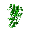
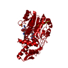
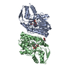
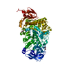
| Title | GDP 4-keto-6-deoxy-D-mannose epimerase reductase S107A | ||||||
 Components Components | GDP-FUCOSE SYNTHETASE | ||||||
 Keywords Keywords | EPIMERASE/REDUCTASE / SDR / RED / EPIMERASE-REDUCTASE complex | ||||||
| Function / homology |  Function and homology information Function and homology informationGDP-L-fucose synthase / GDP-L-fucose synthase activity / 'de novo' GDP-L-fucose biosynthetic process / colanic acid biosynthetic process / NADP+ binding / isomerase activity / protein homodimerization activity / cytoplasm Similarity search - Function | ||||||
| Biological species |  | ||||||
| Method |  X-RAY DIFFRACTION / SYNCHROTRON / MOLECULAR REPLACEMENT / Resolution: 1.6 Å X-RAY DIFFRACTION / SYNCHROTRON / MOLECULAR REPLACEMENT / Resolution: 1.6 Å | ||||||
 Authors Authors | Rosano, C. / Izzo, G. / Bolognesi, M. | ||||||
 Citation Citation | Journal: J.Mol.Biol. / Year: 2000 Title: Probing the Catalytic Mechanism of Gdp-4-Keto-6-Deoxy-D-Mannose Epimerase/Reductase by Kinetic and Crystallographic Characterization of Site-Specific Mutants Authors: Rosano, C. / Bisso, A. / Izzo, G. / Tonetti, M. / Sturla, L. / De Flora, A. / Bolognesi, M. | ||||||
| History |
|
- Structure visualization
| Structure viewer | Molecule: MolmilJmol/JSmol |
|---|
- Downloads & links
-Download
| PDBx/mmCIF format | 1e7q.cif.gz | 88.4 KB | Display | PDBx/mmCIF format |
|---|---|---|---|---|
| PDB format | pdb1e7q.ent.gz | 64.5 KB | Display | PDB format |
| PDBx/mmJSON format | 1e7q.json.gz | Tree view | PDBx/mmJSON format | |
| Others |  Other downloads Other downloads |
-Validation report
| Arichive directory | https://data.pdbj.org/pub/pdb/validation_reports/e7/1e7qftp://data.pdbj.org/pub/pdb/validation_reports/e7/1e7q | HTTPS FTP |
|---|
-Related structure data
| Related structure data |  1e6uC  1e7rC  1e7sC  1bwsS S: Starting model for refinement C: citing same article ( |
|---|---|
| Similar structure data |


-Links
 PDBj
PDBj- Assembly
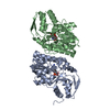
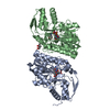
| Deposited unit | 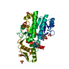
| ||||||||
|---|---|---|---|---|---|---|---|---|---|
| 1 | 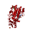
| ||||||||
| Unit cell |
|
-Components
-Protein , 1 types, 1 molecules A
| #1: Protein | Mass: 36128.078 Da / Num. of mol.: 1 / Mutation: YES Source method: isolated from a genetically manipulated source Details: UNKNOWN MOLECULE LABELED AS ACETYLPHOSPHATE / Source: (gene. exp.) References: UniProt: P32055, Isomerases; Racemases and epimerases; Acting on carbohydrates and derivatives |
|---|
-Non-polymers , 5 types, 321 molecules 


| #2: Chemical | ChemComp-NAP /  Mass: 743.405 Da / Num. of mol.: 1 / Source method: obtained synthetically / Formula: C21H28N7O17P3 Mass: 743.405 Da / Num. of mol.: 1 / Source method: obtained synthetically / Formula: C21H28N7O17P3 | ||||
|---|---|---|---|---|---|
| #3: Chemical | ChemComp-UVW /  Mass: 140.032 Da / Num. of mol.: 1 / Source method: obtained synthetically / Formula: C2H5O5P Mass: 140.032 Da / Num. of mol.: 1 / Source method: obtained synthetically / Formula: C2H5O5P | ||||
| #4: Chemical | ChemComp-SO4 /  Mass: 96.063 Da / Num. of mol.: 4 / Source method: obtained synthetically / Formula: SO4 Mass: 96.063 Da / Num. of mol.: 4 / Source method: obtained synthetically / Formula: SO4#5: Chemical | ChemComp-TRS / |  Mass: 122.143 Da / Num. of mol.: 1 / Source method: obtained synthetically / Formula: C4H12NO3 / Comment: pH buffer*YM Mass: 122.143 Da / Num. of mol.: 1 / Source method: obtained synthetically / Formula: C4H12NO3 / Comment: pH buffer*YM#6: Water | ChemComp-HOH / | Mass: 18.015 Da / Num. of mol.: 314 / Source method: isolated from a natural source / Formula: H2O |
-Details
| Compound details | CHAIN A ENGINEERED MUTATION SER107ALA NADP-DEPENDENT CONVERSION OF GDP-4-DEHYDRO-6-DEOXY-D-MANNOSE ...CHAIN A ENGINEERED |
|---|
-Experimental details
-Experiment
| Experiment | Method: X-RAY DIFFRACTION / Number of used crystals: 1 |
|---|
- Sample preparation
| Crystal | Density Matthews: 3.2 Å3/Da / Density % sol: 61.53 % | ||||||||||||||||||||||||
|---|---|---|---|---|---|---|---|---|---|---|---|---|---|---|---|---|---|---|---|---|---|---|---|---|---|
| Crystal grow | Temperature: 294 K / pH: 6.5 Details: 1.5 M LITHIUM SULPHATE, PH 6.5 0.1M TRIS BUFFER, 21C | ||||||||||||||||||||||||
| Crystal grow | *PLUS Temperature: 21 ℃ / Method: unknown / PH range low: 7.8 / PH range high: 6.5 | ||||||||||||||||||||||||
| Components of the solutions | *PLUS
|
-Data collection
| Diffraction | Mean temperature: 100 K |
|---|---|
| Diffraction source | Source: SYNCHROTRON / Site: EMBL/DESY, HAMBURG  / Beamline: BW7A / Wavelength: 0.844 / Beamline: BW7A / Wavelength: 0.844 |
| Detector | Type: MARRESEARCH |
| Radiation | Protocol: SINGLE WAVELENGTH / Monochromatic (M) / Laue (L): M / Scattering type: x-ray |
| Radiation wavelength | Wavelength: 0.844 Å / Relative weight: 1 |
| Reflection | Resolution: 1.6→100 Å / Num. obs: 61196 / % possible obs: 99.3 % / Redundancy: 6.2 % / Rmerge(I) obs: 0.043 / Net I/σ(I): 9.65 |
| Reflection shell | Resolution: 1.6→1.63 Å / Mean I/σ(I) obs: 2.4 / % possible all: 99.1 |
| Reflection shell | *PLUS % possible obs: 99.1 % |
- Processing
| Software |
| ||||||||||||||||||||||||||||||||||||||||||||||||||||||||||||||||||||||||||||||||||||
|---|---|---|---|---|---|---|---|---|---|---|---|---|---|---|---|---|---|---|---|---|---|---|---|---|---|---|---|---|---|---|---|---|---|---|---|---|---|---|---|---|---|---|---|---|---|---|---|---|---|---|---|---|---|---|---|---|---|---|---|---|---|---|---|---|---|---|---|---|---|---|---|---|---|---|---|---|---|---|---|---|---|---|---|---|---|
| Refinement | Method to determine structure: MOLECULAR REPLACEMENT Starting model: PDB ENTRY 1BWS Resolution: 1.6→10 Å / SU B: 1.13 / SU ML: 0.04 / σ(F): 0 / ESU R: 0.068 / ESU R Free: 0.07
| ||||||||||||||||||||||||||||||||||||||||||||||||||||||||||||||||||||||||||||||||||||
| Refinement step | Cycle: LAST / Resolution: 1.6→10 Å
| ||||||||||||||||||||||||||||||||||||||||||||||||||||||||||||||||||||||||||||||||||||
| Refine LS restraints |
|
