 Movie
Movie Controller

[English] 日本語
 Yorodumi
- PDB-2ya0: Catalytic Module of the Multi-modular glycogen-degrading pneumoco... -
+ Open data

- Basic information
| Entry | Database: PDB / ID: 2ya0 | ||||||
|---|---|---|---|---|---|---|---|
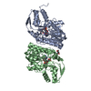
| Title | Catalytic Module of the Multi-modular glycogen-degrading pneumococcal virulence factor SpuA | ||||||
 Components Components | PUTATIVE ALKALINE AMYLOPULLULANASE | ||||||
 Keywords Keywords | HYDROLASE / GLYCOSIDE HYDROLASE | ||||||
| Function / homology |  Function and homology information Function and homology informationlimit dextrinase activity / pullulan binding / amylopectin binding / glycogen binding / alpha-glucan biosynthetic process / pullulanase / pullulanase activity / polysaccharide binding / calcium ion binding / cell surface / extracellular region Similarity search - Function | ||||||
| Biological species |   STREPTOCOCCUS PNEUMONIAE (bacteria) STREPTOCOCCUS PNEUMONIAE (bacteria) | ||||||
| Method |  X-RAY DIFFRACTION / MOLECULAR REPLACEMENT / Resolution: 1.85 Å X-RAY DIFFRACTION / MOLECULAR REPLACEMENT / Resolution: 1.85 Å | ||||||
 Authors Authors | Lammerts van Bueren, A. / Ficko-Blean, E. / Pluvinage, B. / Hehemann, J.H. / Higgins, M.A. / Deng, L. / Ogunniyi, A.D. / Stroeher, U.H. / Warry, N.E. / Burke, R.D. ...Lammerts van Bueren, A. / Ficko-Blean, E. / Pluvinage, B. / Hehemann, J.H. / Higgins, M.A. / Deng, L. / Ogunniyi, A.D. / Stroeher, U.H. / Warry, N.E. / Burke, R.D. / Czjzek, M. / Paton, J.C. / Vocadlo, D.J. / Boraston, A.B. | ||||||
 Citation Citation | Journal: Structure / Year: 2011 Title: The Conformation and Function of a Multimodular Glycogen-Degrading Pneumococcal Virulence Factor. Authors: Lammerts Van Bueren, A. / Ficko-Blean, E. / Pluvinage, B. / Hehemann, J. / Higgins, M.A. / Deng, L. / Ogunniyi, A.D. / Stroeher, U.H. / El Warry, N. / Burke, R.D. / Czjzek, M. / Paton, J.C. ...Authors: Lammerts Van Bueren, A. / Ficko-Blean, E. / Pluvinage, B. / Hehemann, J. / Higgins, M.A. / Deng, L. / Ogunniyi, A.D. / Stroeher, U.H. / El Warry, N. / Burke, R.D. / Czjzek, M. / Paton, J.C. / Vocadlo, D.J. / Boraston, A.B. | ||||||
| History |
| ||||||
| Remark 700 | SHEET DETERMINATION METHOD: DSSP THE SHEETS PRESENTED AS "AE" IN EACH CHAIN ON SHEET RECORDS BELOW ... SHEET DETERMINATION METHOD: DSSP THE SHEETS PRESENTED AS "AE" IN EACH CHAIN ON SHEET RECORDS BELOW IS ACTUALLY AN 8-STRANDED BARREL THIS IS REPRESENTED BY A 9-STRANDED SHEET IN WHICH THE FIRST AND LAST STRANDS ARE IDENTICAL. |
- Structure visualization
| Structure viewer | Molecule: MolmilJmol/JSmol |
|---|
- Downloads & links
-Download
| PDBx/mmCIF format | 2ya0.cif.gz | 175 KB | Display | PDBx/mmCIF format |
|---|---|---|---|---|
| PDB format | pdb2ya0.ent.gz | 135.9 KB | Display | PDB format |
| PDBx/mmJSON format | 2ya0.json.gz | Tree view | PDBx/mmJSON format | |
| Others |  Other downloads Other downloads |
-Validation report
| Arichive directory | https://data.pdbj.org/pub/pdb/validation_reports/ya/2ya0ftp://data.pdbj.org/pub/pdb/validation_reports/ya/2ya0 | HTTPS FTP |
|---|
-Related structure data
| Related structure data |  2ya1C  2ya2C  2fhfS C: citing same article ( S: Starting model for refinement |
|---|---|
| Similar structure data |

-Links
 PDBj
PDBj

- Assembly
| Deposited unit | 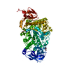
| ||||||||
|---|---|---|---|---|---|---|---|---|---|
| 1 |
| ||||||||
| Unit cell |
|
-Components
| #1: Protein | Mass: 80443.688 Da / Num. of mol.: 1 / Fragment: CATALYTIC DOMAIN, RESIDUES 437-1150 Source method: isolated from a genetically manipulated source Source: (gene. exp.) STREPTOCOCCUS PNEUMONIAE (bacteria) / Strain: TIGR4 / Production host: | ||||
|---|---|---|---|---|---|
| #2: Chemical | ChemComp-CA /   Mass: 40.078 Da / Num. of mol.: 1 / Source method: obtained synthetically / Formula: Ca Mass: 40.078 Da / Num. of mol.: 1 / Source method: obtained synthetically / Formula: Ca | ||||
| #3: Chemical | ChemComp-GOL /   Mass: 92.094 Da / Num. of mol.: 4 / Source method: obtained synthetically / Formula: C3H8O3 Mass: 92.094 Da / Num. of mol.: 4 / Source method: obtained synthetically / Formula: C3H8O3#4: Chemical |   Mass: 22.990 Da / Num. of mol.: 2 / Source method: obtained synthetically / Formula: Na Mass: 22.990 Da / Num. of mol.: 2 / Source method: obtained synthetically / Formula: Na#5: Water | ChemComp-HOH / |  Mass: 18.015 Da / Num. of mol.: 838 / Source method: isolated from a natural source / Formula: H2O Mass: 18.015 Da / Num. of mol.: 838 / Source method: isolated from a natural source / Formula: H2O |
-Experimental details
-Experiment
| Experiment | Method: X-RAY DIFFRACTION / Number of used crystals: 1 |
|---|
- Sample preparation
| Crystal | Density Matthews: 2.32 Å3/Da / Density % sol: 47.05 % / Description: NONE |
|---|
-Data collection
| Diffraction | Mean temperature: 120 K |
|---|---|
| Diffraction source | Source: ROTATING ANODE / Type: RIGAKU MICROMAX-002 / Wavelength: 1.5418 |
| Detector | Type: RIGAKU R-AXIS IV / Detector: IMAGE PLATE |
| Radiation | Protocol: SINGLE WAVELENGTH / Monochromatic (M) / Laue (L): M / Scattering type: x-ray |
| Radiation wavelength | Wavelength: 1.5418 Å / Relative weight: 1 |
| Reflection | Resolution: 1.85→20 Å / Num. obs: 57697 / % possible obs: 96.7 % / Observed criterion σ(I): 2 / Redundancy: 3.73 % / Rmerge(I) obs: 0.06 / Net I/σ(I): 10.2 |
| Reflection shell | Resolution: 1.85→1.92 Å / Redundancy: 3.64 % / Rmerge(I) obs: 0.4 / Mean I/σ(I) obs: 2.3 / % possible all: 94.1 |
- Processing
| Software |
| ||||||||||||||||||||||||||||||||||||||||||||||||||||||||||||||||||||||||||||||||||||||||||||||||||||||||||||||||||||||||||||||||||||||||||||||||||||||||||||||||||||||||||||||||||||||
|---|---|---|---|---|---|---|---|---|---|---|---|---|---|---|---|---|---|---|---|---|---|---|---|---|---|---|---|---|---|---|---|---|---|---|---|---|---|---|---|---|---|---|---|---|---|---|---|---|---|---|---|---|---|---|---|---|---|---|---|---|---|---|---|---|---|---|---|---|---|---|---|---|---|---|---|---|---|---|---|---|---|---|---|---|---|---|---|---|---|---|---|---|---|---|---|---|---|---|---|---|---|---|---|---|---|---|---|---|---|---|---|---|---|---|---|---|---|---|---|---|---|---|---|---|---|---|---|---|---|---|---|---|---|---|---|---|---|---|---|---|---|---|---|---|---|---|---|---|---|---|---|---|---|---|---|---|---|---|---|---|---|---|---|---|---|---|---|---|---|---|---|---|---|---|---|---|---|---|---|---|---|---|---|
| Refinement | Method to determine structure: MOLECULAR REPLACEMENT Starting model: PDB ENTRY 2FHF Resolution: 1.85→20 Å / Cor.coef. Fo:Fc: 0.97 / Cor.coef. Fo:Fc free: 0.946 / SU B: 3.406 / SU ML: 0.1 / Cross valid method: THROUGHOUT / ESU R: 0.15 / ESU R Free: 0.146 / Stereochemistry target values: MAXIMUM LIKELIHOOD Details: HYDROGENS HAVE BEEN ADDED IN THE RIDING POSITIONS. U VALUES REFINED INDIVIDUALLY.
| ||||||||||||||||||||||||||||||||||||||||||||||||||||||||||||||||||||||||||||||||||||||||||||||||||||||||||||||||||||||||||||||||||||||||||||||||||||||||||||||||||||||||||||||||||||||
| Solvent computation | Ion probe radii: 0.8 Å / Shrinkage radii: 0.8 Å / VDW probe radii: 1.4 Å / Solvent model: MASK | ||||||||||||||||||||||||||||||||||||||||||||||||||||||||||||||||||||||||||||||||||||||||||||||||||||||||||||||||||||||||||||||||||||||||||||||||||||||||||||||||||||||||||||||||||||||
| Displacement parameters | Biso mean: 27.763 Å2
| ||||||||||||||||||||||||||||||||||||||||||||||||||||||||||||||||||||||||||||||||||||||||||||||||||||||||||||||||||||||||||||||||||||||||||||||||||||||||||||||||||||||||||||||||||||||
| Refinement step | Cycle: LAST / Resolution: 1.85→20 Å
| ||||||||||||||||||||||||||||||||||||||||||||||||||||||||||||||||||||||||||||||||||||||||||||||||||||||||||||||||||||||||||||||||||||||||||||||||||||||||||||||||||||||||||||||||||||||
| Refine LS restraints |
|