 Movie
Movie Controller

[English] 日本語
 Yorodumi
- PDB-1e30: Crystal structure of the Met148Gln mutant of rusticyanin at 1.5 A... -
+ Open data

- Basic information
| Entry | Database: PDB / ID: 1.0E+30 | ||||||
|---|---|---|---|---|---|---|---|
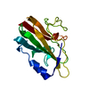
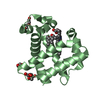
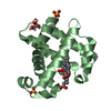
| Title | Crystal structure of the Met148Gln mutant of rusticyanin at 1.5 Angstrom resolution | ||||||
 Components Components | RUSTICYANIN | ||||||
 Keywords Keywords | RUSTICYANIN / MUTANT / AXIAL LIGAND / CUPREDOXIN | ||||||
| Function / homology |  Function and homology information Function and homology information | ||||||
| Biological species |  THIOBACILLUS FERROOXIDANS (bacteria) THIOBACILLUS FERROOXIDANS (bacteria) | ||||||
| Method |  X-RAY DIFFRACTION / SYNCHROTRON / MOLECULAR REPLACEMENT / Resolution: 1.5 Å X-RAY DIFFRACTION / SYNCHROTRON / MOLECULAR REPLACEMENT / Resolution: 1.5 Å | ||||||
 Authors Authors | Hough, M.A. / Strange, R.W. / Hasnain, S.S. | ||||||
 Citation Citation | Journal: Acta Crystallogr.,Sect.D / Year: 2001 Title: Structure of the M148Q mutant of rusticyanin at 1.5 A: a model for the copper site of stellacyanin. Authors: Hough, M.A. / Hall, J.F. / Kanbi, L.D. / Hasnain, S.S. #1: Journal: Biochemistry / Year: 1999 Title: Role of the Axial Ligand in Type 1 Cu Centers Studied by Point Mutations of met148 in Rusticyanin Authors: Hall, J.F. / Kanbi, L.D. / Strange, R.W. / Hasnain, S.S. | ||||||
| History |
|
- Structure visualization
| Structure viewer | Molecule: MolmilJmol/JSmol |
|---|
- Downloads & links
-Download
| PDBx/mmCIF format | 1e30.cif.gz | 72.4 KB | Display | PDBx/mmCIF format |
|---|---|---|---|---|
| PDB format | pdb1e30.ent.gz | 53.8 KB | Display | PDB format |
| PDBx/mmJSON format | 1e30.json.gz | Tree view | PDBx/mmJSON format | |
| Others |  Other downloads Other downloads |
-Validation report
| Summary document | 1e30_validation.pdf.gz | 372.3 KB | Display | wwPDB validaton report |
|---|---|---|---|---|
| Full document | 1e30_full_validation.pdf.gz | 376 KB | Display | |
| Data in XML | 1e30_validation.xml.gz | 7.6 KB | Display | |
| Data in CIF | 1e30_validation.cif.gz | 12.4 KB | Display | |
| Arichive directory | https://data.pdbj.org/pub/pdb/validation_reports/e3/1e30ftp://data.pdbj.org/pub/pdb/validation_reports/e3/1e30 | HTTPS FTP |
-Related structure data
| Similar structure data |
|---|


-Links
 PDBj
PDBj- Assembly
| Deposited unit | 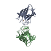
| ||||||||
|---|---|---|---|---|---|---|---|---|---|
| 1 | 
| ||||||||
| 2 | 
| ||||||||
| Unit cell |
|
-Components
| #1: Protein | Mass: 16565.869 Da / Num. of mol.: 2 / Mutation: YES Source method: isolated from a genetically manipulated source Source: (gene. exp.) THIOBACILLUS FERROOXIDANS (bacteria) / Production host: #2: Chemical |   Mass: 63.546 Da / Num. of mol.: 2 / Source method: obtained synthetically / Formula: Cu Mass: 63.546 Da / Num. of mol.: 2 / Source method: obtained synthetically / Formula: Cu#3: Water | ChemComp-HOH / |  Mass: 18.015 Da / Num. of mol.: 179 / Source method: isolated from a natural source / Formula: H2O Mass: 18.015 Da / Num. of mol.: 179 / Source method: isolated from a natural source / Formula: H2OCompound details | CHAIN A, B ENGINEERED | Has protein modification | N | |
|---|
-Experimental details
-Experiment
| Experiment | Method: X-RAY DIFFRACTION / Number of used crystals: 1 |
|---|
- Sample preparation
| Crystal | Density Matthews: 2.1 Å3/Da / Density % sol: 41.3 % | ||||||||||||||||||||||||||||||||||||
|---|---|---|---|---|---|---|---|---|---|---|---|---|---|---|---|---|---|---|---|---|---|---|---|---|---|---|---|---|---|---|---|---|---|---|---|---|---|
| Crystal grow | Method: vapor diffusion / pH: 4 Details: 30% PEG-8000, 100MM MES, 50MM CITRIC ACID, PH 4.0 VAPOUR DIFFUSION. PROTEIN CONCENTRATION 7 MG./ML | ||||||||||||||||||||||||||||||||||||
| Crystal grow | *PLUS Temperature: 293 K / Method: vapor diffusion, hanging dropDetails: drop was mixed with an equal volume of reservoir solution | ||||||||||||||||||||||||||||||||||||
| Components of the solutions | *PLUS
|
-Data collection
| Diffraction | Mean temperature: 100 K |
|---|---|
| Diffraction source | Source: SYNCHROTRON / Site: Photon Factory  / Beamline: BL-6A / Wavelength: 1 / Beamline: BL-6A / Wavelength: 1 |
| Detector | Type: FUJI / Detector: IMAGE PLATE / Date: Apr 15, 2000 |
| Radiation | Protocol: SINGLE WAVELENGTH / Monochromatic (M) / Laue (L): M / Scattering type: x-ray |
| Radiation wavelength | Wavelength: 1 Å / Relative weight: 1 |
| Reflection | Resolution: 1.5→52.7 Å / Num. obs: 33706 / % possible obs: 76.5 % / Redundancy: 2.1 % / Biso Wilson estimate: 11 Å2 / Rmerge(I) obs: 0.056 / Rsym value: 0.056 / Net I/σ(I): 12 |
| Reflection shell | Resolution: 1.5→1.53 Å / Redundancy: 2 % / Rmerge(I) obs: 0.203 / Mean I/σ(I) obs: 3.1 / Rsym value: 0.203 / % possible all: 70.7 |
| Reflection | *PLUS Num. measured all: 62663 |
| Reflection shell | *PLUS % possible obs: 70.7 % / Mean I/σ(I) obs: 3 |
- Processing
| Software |
| ||||||||||||||||||||||||||||||||||||||||||||||||||||||||||||||||||||||||||||||||||||
|---|---|---|---|---|---|---|---|---|---|---|---|---|---|---|---|---|---|---|---|---|---|---|---|---|---|---|---|---|---|---|---|---|---|---|---|---|---|---|---|---|---|---|---|---|---|---|---|---|---|---|---|---|---|---|---|---|---|---|---|---|---|---|---|---|---|---|---|---|---|---|---|---|---|---|---|---|---|---|---|---|---|---|---|---|---|
| Refinement | Method to determine structure: MOLECULAR REPLACEMENT / Resolution: 1.5→52.7 Å / SU B: 1.60728 / SU ML: 0.06 / Cross valid method: THROUGHOUT / σ(F): 0 / ESU R: 0.1 / ESU R Free: 0.1 Details: THE TWO N-TERMINAL RESIDUES ARE NOT SEEN IN ELECTRON DENSITY
| ||||||||||||||||||||||||||||||||||||||||||||||||||||||||||||||||||||||||||||||||||||
| Displacement parameters | Biso mean: 11 Å2 | ||||||||||||||||||||||||||||||||||||||||||||||||||||||||||||||||||||||||||||||||||||
| Refinement step | Cycle: LAST / Resolution: 1.5→52.7 Å
| ||||||||||||||||||||||||||||||||||||||||||||||||||||||||||||||||||||||||||||||||||||
| Refine LS restraints |
|
