 Movie
Movie Controller


+ Open data

- Basic information
| Entry | Database: PDB / ID: 1dxk | ||||||
|---|---|---|---|---|---|---|---|
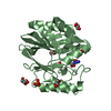
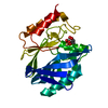
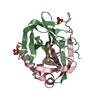
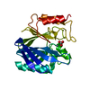
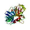
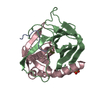
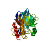
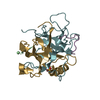
| Title | Metallo-beta-lactamase from Bacillus cereus 569/H/9 C168S mutant | ||||||
 Components Components | CLASS B BETA-LACTAMASE | ||||||
 Keywords Keywords | HYDROLASE / HYDROLASE (BETA-LACTAMASE) / METALLO BETA-LACTAMASE / ZINC | ||||||
| Function / homology |  Function and homology information Function and homology informationantibiotic catabolic process / beta-lactamase activity / beta-lactamase / periplasmic space / response to antibiotic / zinc ion binding Similarity search - Function | ||||||
| Biological species |  | ||||||
| Method |  X-RAY DIFFRACTION / MIR / Resolution: 1.85 Å X-RAY DIFFRACTION / MIR / Resolution: 1.85 Å | ||||||
 Authors Authors | Chantalat, L. / Duee, E. / Dideberg, O. | ||||||
 Citation Citation | Journal: Protein Sci. / Year: 2000 Title: Structural effects of the active site mutation cysteine to serine in Bacillus cereus zinc-beta-lactamase. Authors: Chantalat, L. / Duee, E. / Galleni, M. / Frere, J.M. / Dideberg, O. #1: Journal: Acta Crystallogr.,Sect.D / Year: 1998Title: 1.85 A Resolution Structure of the Zinc(II) Beta-Lactamase from Bacillus Cereus Authors: Carfi, A. / Duee, E. / Galleni, M. / Frere, J.M. / Dideberg, O. #2: Journal: Embo J. / Year: 1995Title: The 3-D Structure of a Zinc Metallo-Beta-Lactamase from Bacillus Cereus Reveals a New Type of Protein Fold Authors: Carfi, A. / Pares, S. / Duee, E. / Galleni, M. / Duez, C. / Frere, J.M. / Dideberg, O. | ||||||
| History |
|
- Structure visualization
| Structure viewer | Molecule: MolmilJmol/JSmol |
|---|
- Downloads & links
-Download
| PDBx/mmCIF format | 1dxk.cif.gz | 62.9 KB | Display | PDBx/mmCIF format |
|---|---|---|---|---|
| PDB format | pdb1dxk.ent.gz | 44.7 KB | Display | PDB format |
| PDBx/mmJSON format | 1dxk.json.gz | Tree view | PDBx/mmJSON format | |
| Others |  Other downloads Other downloads |
-Validation report
| Arichive directory | https://data.pdbj.org/pub/pdb/validation_reports/dx/1dxkftp://data.pdbj.org/pub/pdb/validation_reports/dx/1dxk | HTTPS FTP |
|---|
-Related structure data
| Related structure data |  1bvtS S: Starting model for refinement |
|---|---|
| Similar structure data |


-Links
 PDBj
PDBj


- Assembly
| Deposited unit | 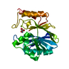
| ||||||||
|---|---|---|---|---|---|---|---|---|---|
| 1 |
| ||||||||
| Unit cell |
|
-Components
| #1: Protein | Mass: 24979.469 Da / Num. of mol.: 1 / Mutation: YES Source method: isolated from a genetically manipulated source Details: 1MM ZN IN THE BUFFER / Source: (gene. exp.) |
|---|---|
| #2: Chemical | ChemComp-ZN /   Mass: 65.409 Da / Num. of mol.: 1 / Source method: obtained synthetically / Formula: Zn Mass: 65.409 Da / Num. of mol.: 1 / Source method: obtained synthetically / Formula: Zn |
| #3: Chemical | ChemComp-BCT /   Mass: 61.017 Da / Num. of mol.: 1 / Source method: obtained synthetically / Formula: CHO3 / Comment: pH buffer*YM Mass: 61.017 Da / Num. of mol.: 1 / Source method: obtained synthetically / Formula: CHO3 / Comment: pH buffer*YM |
| #4: Water | ChemComp-HOH /  Mass: 18.015 Da / Num. of mol.: 232 / Source method: isolated from a natural source / Formula: H2O Mass: 18.015 Da / Num. of mol.: 232 / Source method: isolated from a natural source / Formula: H2O |
-Experimental details
-Experiment
| Experiment | Method: X-RAY DIFFRACTION / Number of used crystals: 1 |
|---|
- Sample preparation
| Crystal | Density Matthews: 2.3 Å3/Da / Density % sol: 38 % | ||||||||||||||||||||||||||||||||||||||||||||||||
|---|---|---|---|---|---|---|---|---|---|---|---|---|---|---|---|---|---|---|---|---|---|---|---|---|---|---|---|---|---|---|---|---|---|---|---|---|---|---|---|---|---|---|---|---|---|---|---|---|---|
| Crystal grow | pH: 5.6 / Details: pH 5.60 | ||||||||||||||||||||||||||||||||||||||||||||||||
| Crystal grow | *PLUS Temperature: 8 ℃ / pH: 6.5 / Method: vapor diffusion, hanging drop | ||||||||||||||||||||||||||||||||||||||||||||||||
| Components of the solutions | *PLUS
|
-Data collection
| Diffraction | Mean temperature: 100 K |
|---|---|
| Diffraction source | Source: ROTATING ANODE / Type: RIGAKU / Wavelength: 1.5418 |
| Detector | Type: MARRESEARCH / Detector: IMAGE PLATE / Date: Nov 15, 1998 / Details: MIRRORS |
| Radiation | Protocol: SINGLE WAVELENGTH / Monochromatic (M) / Laue (L): M / Scattering type: x-ray |
| Radiation wavelength | Wavelength: 1.5418 Å / Relative weight: 1 |
| Reflection | Resolution: 1.85→20 Å / Num. obs: 17009 / % possible obs: 89.9 % / Redundancy: 3.82 % / Biso Wilson estimate: 16.6 Å2 / Rsym value: 0.043 |
| Reflection shell | Resolution: 1.85→1.92 Å / Redundancy: 3.75 % / Rsym value: 0.069 / % possible all: 88.8 |
| Reflection | *PLUS Highest resolution: 1.85 Å / Lowest resolution: 20 Å / Rmerge(I) obs: 0.043 |
| Reflection shell | *PLUS % possible obs: 88.8 % / Num. unique obs: 1648 / Rmerge(I) obs: 0.069 |
- Processing
| Software |
| ||||||||||||||||||||||||||||||||||||||||||||||||||||||||||||
|---|---|---|---|---|---|---|---|---|---|---|---|---|---|---|---|---|---|---|---|---|---|---|---|---|---|---|---|---|---|---|---|---|---|---|---|---|---|---|---|---|---|---|---|---|---|---|---|---|---|---|---|---|---|---|---|---|---|---|---|---|---|
| Refinement | Method to determine structure: MIR Starting model: PDB ENTRY 1BVT Resolution: 1.85→8 Å / Cross valid method: THROUGHOUT / σ(F): 2 Details: DISORDERED RESIDUES, (I.E. FOR WHICH NO COORDINATES ARE REPORTED BUT THE OCCUPANCY IS SET TO ZERO) ARE 11 - 14, 32 - 38, AND GLU 212 HAS TWO CONFORMATIONS
| ||||||||||||||||||||||||||||||||||||||||||||||||||||||||||||
| Displacement parameters | Biso mean: 17.57 Å2 | ||||||||||||||||||||||||||||||||||||||||||||||||||||||||||||
| Refinement step | Cycle: LAST / Resolution: 1.85→8 Å
| ||||||||||||||||||||||||||||||||||||||||||||||||||||||||||||
| Refine LS restraints |
| ||||||||||||||||||||||||||||||||||||||||||||||||||||||||||||
| LS refinement shell | Resolution: 1.85→1.93 Å / Total num. of bins used: 8
| ||||||||||||||||||||||||||||||||||||||||||||||||||||||||||||
| Xplor file | Serial no: 1 / Param file: PARHCSDX.PRO / Topol file: TOPH19X.PRO | ||||||||||||||||||||||||||||||||||||||||||||||||||||||||||||
| Software | *PLUS Name: X-PLOR / Version: 3.1 / Classification: refinement | ||||||||||||||||||||||||||||||||||||||||||||||||||||||||||||
| Refinement | *PLUS Rfactor Rfree: 0.2612 | ||||||||||||||||||||||||||||||||||||||||||||||||||||||||||||
| Solvent computation | *PLUS | ||||||||||||||||||||||||||||||||||||||||||||||||||||||||||||
| Displacement parameters | *PLUS |
