 Movie
Movie Controller


+ Open data

- Basic information
| Entry | Database: PDB / ID: 1bvt | |||||||||
|---|---|---|---|---|---|---|---|---|---|---|
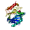
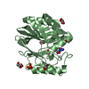
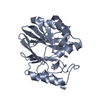
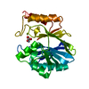
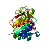
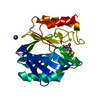
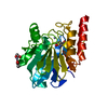
| Title | METALLO-BETA-LACTAMASE FROM BACILLUS CEREUS 569/H/9 | |||||||||
 Components Components | PROTEIN (BETA-LACTAMASE) | |||||||||
 Keywords Keywords | HYDROLASE / HYDROLASE (BETA-LACTAMASE) / METALLO BETA-LACTAMASE / ZINC | |||||||||
| Function / homology |  Function and homology information Function and homology informationantibiotic catabolic process / beta-lactamase activity / beta-lactamase / periplasmic space / response to antibiotic / zinc ion binding Similarity search - Function | |||||||||
| Biological species |  | |||||||||
| Method |  X-RAY DIFFRACTION / SYNCHROTRON / MOLECULAR REPLACEMENT / Resolution: 1.85 Å X-RAY DIFFRACTION / SYNCHROTRON / MOLECULAR REPLACEMENT / Resolution: 1.85 Å | |||||||||
 Authors Authors | Carfi, A. / Duee, E. / Dideberg, O. | |||||||||
 Citation Citation | Journal: Acta Crystallogr.,Sect.D / Year: 1998 Title: 1.85 A resolution structure of the zinc (II) beta-lactamase from Bacillus cereus. Authors: Carfi, A. / Duee, E. / Galleni, M. / Frere, J.M. / Dideberg, O. #1: Journal: Embo J. / Year: 1995Title: The 3-D Structure of a Zinc Metallo-Beta-Lactamase from Bacillus Cereus Reveals a New Type of Protein Fold Authors: Carfi, A. / Pares, S. / Duee, E. / Galleni, M. / Duez, C. / Frere, J.M. / Dideberg, O. | |||||||||
| History |
|
- Structure visualization
| Structure viewer | Molecule: MolmilJmol/JSmol |
|---|
- Downloads & links
-Download
| PDBx/mmCIF format | 1bvt.cif.gz | 60.6 KB | Display | PDBx/mmCIF format |
|---|---|---|---|---|
| PDB format | pdb1bvt.ent.gz | 43.8 KB | Display | PDB format |
| PDBx/mmJSON format | 1bvt.json.gz | Tree view | PDBx/mmJSON format | |
| Others |  Other downloads Other downloads |
-Validation report
| Arichive directory | https://data.pdbj.org/pub/pdb/validation_reports/bv/1bvtftp://data.pdbj.org/pub/pdb/validation_reports/bv/1bvt | HTTPS FTP |
|---|
-Related structure data
| Similar structure data |
|---|


-Links
 PDBj
PDBj


- Assembly
| Deposited unit | 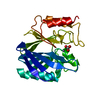
| ||||||||
|---|---|---|---|---|---|---|---|---|---|
| 1 |
| ||||||||
| Unit cell |
|
-Components
| #1: Protein | Mass: 24995.533 Da / Num. of mol.: 1 Source method: isolated from a genetically manipulated source Details: 100 MICROMOLAR ZN IN THE BUFFER / Source: (gene. exp.) | ||||
|---|---|---|---|---|---|
| #2: Chemical |   Mass: 65.409 Da / Num. of mol.: 2 / Source method: obtained synthetically / Formula: Zn Mass: 65.409 Da / Num. of mol.: 2 / Source method: obtained synthetically / Formula: Zn#3: Chemical | ChemComp-BCT / |   Mass: 61.017 Da / Num. of mol.: 1 / Source method: obtained synthetically / Formula: CHO3 / Comment: pH buffer*YM Mass: 61.017 Da / Num. of mol.: 1 / Source method: obtained synthetically / Formula: CHO3 / Comment: pH buffer*YM#4: Water | ChemComp-HOH / |  Mass: 18.015 Da / Num. of mol.: 205 / Source method: isolated from a natural source / Formula: H2O Mass: 18.015 Da / Num. of mol.: 205 / Source method: isolated from a natural source / Formula: H2O |
-Experimental details
-Experiment
| Experiment | Method: X-RAY DIFFRACTION / Number of used crystals: 1 |
|---|
- Sample preparation
| Crystal | Density Matthews: 2.33 Å3/Da / Density % sol: 38 % | ||||||||||||||||||||||||||||||||||||||||||||||||||||||
|---|---|---|---|---|---|---|---|---|---|---|---|---|---|---|---|---|---|---|---|---|---|---|---|---|---|---|---|---|---|---|---|---|---|---|---|---|---|---|---|---|---|---|---|---|---|---|---|---|---|---|---|---|---|---|---|
| Crystal grow | pH: 5.6 / Details: SEE IN ACTA CRYSTALLOG PAPER, pH 5.6 | ||||||||||||||||||||||||||||||||||||||||||||||||||||||
| Crystal | *PLUS | ||||||||||||||||||||||||||||||||||||||||||||||||||||||
| Crystal grow | *PLUS Temperature: 281 K / pH: 6.5 / Method: vapor diffusion, hanging drop | ||||||||||||||||||||||||||||||||||||||||||||||||||||||
| Components of the solutions | *PLUS
|
-Data collection
| Diffraction | Mean temperature: 100 K |
|---|---|
| Diffraction source | Source: SYNCHROTRON / Site: ESRF  / Beamline: BM02 / Wavelength: 0.982 / Beamline: BM02 / Wavelength: 0.982 |
| Detector | Detector: CCD / Date: Feb 15, 1996 |
| Radiation | Protocol: SINGLE WAVELENGTH / Monochromatic (M) / Laue (L): M / Scattering type: x-ray |
| Radiation wavelength | Wavelength: 0.982 Å / Relative weight: 1 |
| Reflection | Resolution: 1.85→20 Å / Num. obs: 15240 / % possible obs: 80.9 % / Observed criterion σ(I): 2 / Redundancy: 2.6 % / Rsym value: 7 |
| Reflection shell | Resolution: 1.85→1.92 Å / Redundancy: 2 % / Rsym value: 10 / % possible all: 79.3 |
| Reflection | *PLUS Rmerge(I) obs: 0.07 |
| Reflection shell | *PLUS % possible obs: 77.7 % / Rmerge(I) obs: 0.104 |
- Processing
| Software |
| ||||||||||||||||||||||||||||||||||||||||||||||||||||||||||||
|---|---|---|---|---|---|---|---|---|---|---|---|---|---|---|---|---|---|---|---|---|---|---|---|---|---|---|---|---|---|---|---|---|---|---|---|---|---|---|---|---|---|---|---|---|---|---|---|---|---|---|---|---|---|---|---|---|---|---|---|---|---|
| Refinement | Method to determine structure: MOLECULAR REPLACEMENT Starting model: B.CEREUS EMBO J. Resolution: 1.85→8 Å / σ(F): 2 Details: FINAL RMS COORD. SHIFT 0.015 ANGSTROMS MEAN B VALUE (MAIN CHAIN, A**2): 13. MEAN B VALUE (SIDE CHAIN, A**2): 15. MEAN B VALUE (SOLVENT, A**2): 26. MEAN B VALUE (ZINC IONS, A**2): 24.
| ||||||||||||||||||||||||||||||||||||||||||||||||||||||||||||
| Refine analyze | Luzzati sigma a obs: 0.2 Å | ||||||||||||||||||||||||||||||||||||||||||||||||||||||||||||
| Refinement step | Cycle: LAST / Resolution: 1.85→8 Å
| ||||||||||||||||||||||||||||||||||||||||||||||||||||||||||||
| Refine LS restraints |
| ||||||||||||||||||||||||||||||||||||||||||||||||||||||||||||
| Software | *PLUS Name: X-PLOR / Version: 3.1 / Classification: refinement | ||||||||||||||||||||||||||||||||||||||||||||||||||||||||||||
| Refinement | *PLUS Lowest resolution: 8 Å / σ(F): 2 / % reflection Rfree: 5 % / Rfactor obs: 0.214 | ||||||||||||||||||||||||||||||||||||||||||||||||||||||||||||
| Solvent computation | *PLUS | ||||||||||||||||||||||||||||||||||||||||||||||||||||||||||||
| Displacement parameters | *PLUS | ||||||||||||||||||||||||||||||||||||||||||||||||||||||||||||
| Refine LS restraints | *PLUS
|
