 Movie
Movie Controller


+ Open data

- Basic information
| Entry | Database: PDB / ID: 1dwr | ||||||
|---|---|---|---|---|---|---|---|
| Title | MYOGLOBIN (HORSE HEART) WILD-TYPE COMPLEXED WITH CO | ||||||
 Components Components | Myoglobin | ||||||
 Keywords Keywords | OXYGEN TRANSPORT / RESPIRATORY PROTEIN | ||||||
| Function / homology |  Function and homology information Function and homology informationOxidoreductases; Acting on other nitrogenous compounds as donors / nitrite reductase activity / oxygen transport / sarcoplasm / Oxidoreductases; Acting on a peroxide as acceptor; Peroxidases / skeletal muscle contraction / removal of superoxide radicals / oxygen carrier activity / peroxidase activity / oxygen binding ...Oxidoreductases; Acting on other nitrogenous compounds as donors / nitrite reductase activity / oxygen transport / sarcoplasm / Oxidoreductases; Acting on a peroxide as acceptor; Peroxidases / skeletal muscle contraction / removal of superoxide radicals / oxygen carrier activity / peroxidase activity / oxygen binding / heme binding / metal ion binding Similarity search - Function | ||||||
| Biological species |  | ||||||
| Method |  X-RAY DIFFRACTION / SYNCHROTRON / MOLECULAR REPLACEMENT / Resolution: 1.45 Å X-RAY DIFFRACTION / SYNCHROTRON / MOLECULAR REPLACEMENT / Resolution: 1.45 Å | ||||||
 Authors Authors | Chu, K. / Vojtechovsky, J. / McMahon, B.H. / Sweet, R.M. / Berendzen, J. / Schlichting, I. | ||||||
 Citation Citation | Journal: Nature / Year: 2000 Title: Crystal Structure of a New Ligand Binding Intermediate in Wildtype Carbonmonoxy Myoglobin Authors: Chu, K. / Vojtechovsky, J. / Mcmahon, B.H. / Sweet, R.M. / Berendzen, J. / Schlichting, I. #1: Journal: Biochim.Biophys.Acta / Year: 1997Title: A Myoglobin Variant with a Polar Substitution in a Conserved Hydrophobic Cluster in the Heme Binding Pocket Authors: Maurus, R. / Overall, C.M. / Bogumil, R. / Luo, Y. / Mauk, A.G. / Smith, M. / Brayer, G.D. #2: Journal: Nature / Year: 1994Title: Crystal Structure of Photolysed Myoglobin Authors: Schlichting, I. / Berendzen, J. / Phillips Jr, G.N. / Sweet, R.M. #3: Journal: J.Mol.Biol. / Year: 1987 Title: Crystallization and Preliminary Diffraction Data for Horse Heart Metmyoglobin Authors: Sherwood, C. / Mauk, A.G. / Brayer, G.D. | ||||||
| History |
|
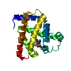
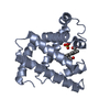
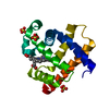
- Structure visualization
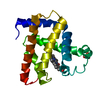
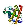
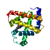
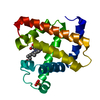
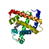
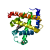
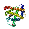
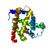
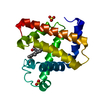
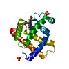
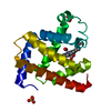
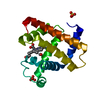
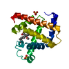
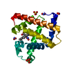
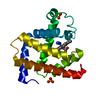
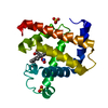
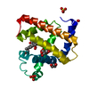
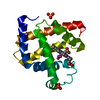
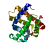
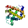
| Structure viewer | Molecule: MolmilJmol/JSmol |
|---|
- Downloads & links
-Download
| PDBx/mmCIF format | 1dwr.cif.gz | 49.1 KB | Display | PDBx/mmCIF format |
|---|---|---|---|---|
| PDB format | pdb1dwr.ent.gz | 33.2 KB | Display | PDB format |
| PDBx/mmJSON format | 1dwr.json.gz | Tree view | PDBx/mmJSON format | |
| Others |  Other downloads Other downloads |
-Validation report
| Arichive directory | https://data.pdbj.org/pub/pdb/validation_reports/dw/1dwrftp://data.pdbj.org/pub/pdb/validation_reports/dw/1dwr | HTTPS FTP |
|---|
-Related structure data
| Related structure data | 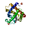 1dwsC 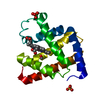 1dwtC 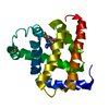 1aziS C: citing same article ( S: Starting model for refinement |
|---|---|
| Similar structure data |


-Links
 PDBj
PDBj


- Assembly
| Deposited unit | 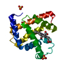
| ||||||||
|---|---|---|---|---|---|---|---|---|---|
| 1 |
| ||||||||
| Unit cell |
| ||||||||
| Details | BIOLOGICAL_UNIT: MONOMER |
-Components
| #1: Protein | Mass: 16983.514 Da / Num. of mol.: 1 / Source method: isolated from a natural source / Source: (natural) | ||
|---|---|---|---|
| #2: Chemical | ChemComp-HEM /   Mass: 616.487 Da / Num. of mol.: 1 / Source method: obtained synthetically / Formula: C34H32FeN4O4 Mass: 616.487 Da / Num. of mol.: 1 / Source method: obtained synthetically / Formula: C34H32FeN4O4 | ||
| #3: Chemical | ChemComp-CMO /   Mass: 28.010 Da / Num. of mol.: 1 / Source method: obtained synthetically / Formula: CO Mass: 28.010 Da / Num. of mol.: 1 / Source method: obtained synthetically / Formula: CO | ||
| #4: Chemical |   Mass: 96.063 Da / Num. of mol.: 2 / Source method: obtained synthetically / Formula: SO4 Mass: 96.063 Da / Num. of mol.: 2 / Source method: obtained synthetically / Formula: SO4#5: Water | ChemComp-HOH / |  Mass: 18.015 Da / Num. of mol.: 132 / Source method: isolated from a natural source / Formula: H2O Mass: 18.015 Da / Num. of mol.: 132 / Source method: isolated from a natural source / Formula: H2O |
-Experimental details
-Experiment
| Experiment | Method: X-RAY DIFFRACTION / Number of used crystals: 1 |
|---|
- Sample preparation
| Crystal | Density Matthews: 1.84 Å3/Da / Density % sol: 33.18 % | ||||||||||||||||||||||||||||||
|---|---|---|---|---|---|---|---|---|---|---|---|---|---|---|---|---|---|---|---|---|---|---|---|---|---|---|---|---|---|---|---|
| Crystal grow | Method: vapor diffusion, hanging drop / pH: 7.5 Details: HHMB(SIGMA) WAS CRYSTALLIZED AT ROOM TEMPERATURE BY EQUILIBRATING 10 UL DROPS OF 5 MG/ML PROTEIN IN 1.7-1.8 M AMMONIUM SULFATE, 0.1 M TRIS HCL PH 7.5 USING THE HANGING DROP GEOMETRY | ||||||||||||||||||||||||||||||
| Crystal grow | *PLUS Method: vapor diffusion, hanging drop | ||||||||||||||||||||||||||||||
| Components of the solutions | *PLUS
|
-Data collection
| Diffraction | Mean temperature: 100 K |
|---|---|
| Diffraction source | Source: SYNCHROTRON / Site: NSLS  / Beamline: X12C / Wavelength: 1.1 / Beamline: X12C / Wavelength: 1.1 |
| Detector | Type: PRINCETON SCIENTIFIC / Detector: CCD / Date: Jun 2, 1998 / Details: MIRRORS |
| Radiation | Protocol: SINGLE WAVELENGTH / Monochromatic (M) / Laue (L): M / Scattering type: x-ray |
| Radiation wavelength | Wavelength: 1.1 Å / Relative weight: 1 |
| Reflection | Resolution: 1.4→20 Å / Num. obs: 23795 / % possible obs: 96.1 % / Observed criterion σ(I): 0 / Redundancy: 2.47 % / Rmerge(I) obs: 0.07 / Rsym value: 0.07 / Net I/σ(I): 9.2 |
| Reflection shell | Resolution: 1.4→1.5 Å / Redundancy: 1.6 % / Rmerge(I) obs: 0.227 / Mean I/σ(I) obs: 2.3 / Rsym value: 0.227 / % possible all: 87.2 |
| Reflection shell | *PLUS % possible obs: 87.2 % |
- Processing
| Software |
| ||||||||||||||||||||||||||||||||||||||||||||||||||||||||||||
|---|---|---|---|---|---|---|---|---|---|---|---|---|---|---|---|---|---|---|---|---|---|---|---|---|---|---|---|---|---|---|---|---|---|---|---|---|---|---|---|---|---|---|---|---|---|---|---|---|---|---|---|---|---|---|---|---|---|---|---|---|---|
| Refinement | Method to determine structure: MOLECULAR REPLACEMENT Starting model: 1AZI Resolution: 1.45→20 Å / Cross valid method: THROUGHOUT / σ(F): 0 / Details: LYS47 AND SER92 HAVE ALTERNATE CONFORMATIONS
| ||||||||||||||||||||||||||||||||||||||||||||||||||||||||||||
| Refinement step | Cycle: LAST / Resolution: 1.45→20 Å
| ||||||||||||||||||||||||||||||||||||||||||||||||||||||||||||
| Refine LS restraints |
|
