 Movie
Movie Controller

[English] 日本語
 Yorodumi
- PDB-1doj: Crystal structure of human alpha-thrombin*RWJ-51438 complex at 1.7 A -
+ Open data

- Basic information
| Entry | Database: PDB / ID: 1doj | ||||||
|---|---|---|---|---|---|---|---|
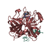
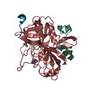
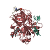
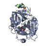
| Title | Crystal structure of human alpha-thrombin*RWJ-51438 complex at 1.7 A | ||||||
 Components Components |
| ||||||
 Keywords Keywords | HYDROLASE/HYDROLASE INHIBITOR / thrombin / serine protease / enzyme inhibition / HYDROLASE-HYDROLASE INHIBITOR COMPLEX | ||||||
| Function / homology |  Function and homology information Function and homology informationcytolysis by host of symbiont cells / thrombospondin receptor activity / thrombin-activated receptor signaling pathway / Defective factor XII causes hereditary angioedema / thrombin / negative regulation of astrocyte differentiation / regulation of blood coagulation / positive regulation of phospholipase C-activating G protein-coupled receptor signaling pathway / neutrophil-mediated killing of gram-negative bacterium / Defective F8 cleavage by thrombin ...cytolysis by host of symbiont cells / thrombospondin receptor activity / thrombin-activated receptor signaling pathway / Defective factor XII causes hereditary angioedema / thrombin / negative regulation of astrocyte differentiation / regulation of blood coagulation / positive regulation of phospholipase C-activating G protein-coupled receptor signaling pathway / neutrophil-mediated killing of gram-negative bacterium / Defective F8 cleavage by thrombin / Platelet Aggregation (Plug Formation) / ligand-gated ion channel signaling pathway / positive regulation of collagen biosynthetic process / negative regulation of platelet activation / negative regulation of blood coagulation / positive regulation of blood coagulation / negative regulation of fibrinolysis / regulation of cytosolic calcium ion concentration / Transport of gamma-carboxylated protein precursors from the endoplasmic reticulum to the Golgi apparatus / Gamma-carboxylation of protein precursors / Common Pathway of Fibrin Clot Formation / Removal of aminoterminal propeptides from gamma-carboxylated proteins / fibrinolysis / Intrinsic Pathway of Fibrin Clot Formation / negative regulation of proteolysis / negative regulation of cytokine production involved in inflammatory response / positive regulation of release of sequestered calcium ion into cytosol / Peptide ligand-binding receptors / Regulation of Complement cascade / acute-phase response / Cell surface interactions at the vascular wall / positive regulation of receptor signaling pathway via JAK-STAT / lipopolysaccharide binding / growth factor activity / serine-type endopeptidase inhibitor activity / positive regulation of insulin secretion / platelet activation / response to wounding / Golgi lumen / positive regulation of protein localization to nucleus / Regulation of Insulin-like Growth Factor (IGF) transport and uptake by Insulin-like Growth Factor Binding Proteins (IGFBPs) / positive regulation of reactive oxygen species metabolic process / blood coagulation / antimicrobial humoral immune response mediated by antimicrobial peptide / regulation of cell shape / heparin binding / Thrombin signalling through proteinase activated receptors (PARs) / : / positive regulation of cell growth / blood microparticle / G alpha (q) signalling events / cell surface receptor signaling pathway / positive regulation of phosphatidylinositol 3-kinase/protein kinase B signal transduction / receptor ligand activity / endoplasmic reticulum lumen / signaling receptor binding / serine-type endopeptidase activity / positive regulation of cell population proliferation / calcium ion binding / proteolysis / extracellular space / extracellular exosome / extracellular region / plasma membrane Similarity search - Function | ||||||
| Biological species |  Homo sapiens (human) Homo sapiens (human) | ||||||
| Method |  X-RAY DIFFRACTION / Resolution: 1.7 Å X-RAY DIFFRACTION / Resolution: 1.7 Å | ||||||
 Authors Authors | Recacha, R. / Costanzo, M.J. / Maryanoff, B.E. / Carson, M. / DeLucas, L. / Chattopadhyay, D. | ||||||
 Citation Citation | Journal: Acta Crystallogr.,Sect.D / Year: 2000 Title: Structure of human alpha-thrombin complexed with RWJ-51438 at 1.7 A: unusual perturbation of the 60A-60I insertion loop. Authors: Recacha, R. / Costanzo, M.J. / Maryanoff, B.E. / Carson, M. / DeLucas, L. / Chattopadhyay, D. #1: Journal: Biochemistry / Year: 1992Title: Structure of the Hirulog 3-Thrombin Complex and Nature of the S' Subsites of Substrates and Inhibitors Authors: Qiu, X. / Padmanabhan, K.P. / Carperos, V.E. / Tulinsky, A. / Kline, T. / Maraganore, J.M. / Fenton II, J.W. #2: Journal: Biophys.J. / Year: 1996Title: Crystal Structure of Thrombin with Thiazole-Containing Inhibitors: Probes of the S1' Binding Site Authors: Matthews, J.H. / Krishnan, R. / Costanzo, M.J. / Maryanoff, B.E. / Tulinsky, A. #3: Journal: Protein Sci. / Year: 1992Title: The Refined 1.9-A X-ray Crystal Structure of D-Pro-Phe-Arg Chloromethylketone-Inhibited Human Alpha-Thrombin: Structure Analysis, Overall Structure, Electrostatic Properties, Detailed Active- ...Title: The Refined 1.9-A X-ray Crystal Structure of D-Pro-Phe-Arg Chloromethylketone-Inhibited Human Alpha-Thrombin: Structure Analysis, Overall Structure, Electrostatic Properties, Detailed Active-Site Geometry, and Structure-Function Relationships Authors: Bode, W. / Turk, D. / Karshikov, A. | ||||||
| History |
|
- Structure visualization
| Structure viewer | Molecule: MolmilJmol/JSmol |
|---|
- Downloads & links
-Download
| PDBx/mmCIF format | 1doj.cif.gz | 88.7 KB | Display | PDBx/mmCIF format |
|---|---|---|---|---|
| PDB format | pdb1doj.ent.gz | 63.7 KB | Display | PDB format |
| PDBx/mmJSON format | 1doj.json.gz | Tree view | PDBx/mmJSON format | |
| Others |  Other downloads Other downloads |
-Validation report
| Summary document | 1doj_validation.pdf.gz | 812.1 KB | Display | wwPDB validaton report |
|---|---|---|---|---|
| Full document | 1doj_full_validation.pdf.gz | 819.7 KB | Display | |
| Data in XML | 1doj_validation.xml.gz | 19.2 KB | Display | |
| Data in CIF | 1doj_validation.cif.gz | 28.2 KB | Display | |
| Arichive directory | https://data.pdbj.org/pub/pdb/validation_reports/do/1dojftp://data.pdbj.org/pub/pdb/validation_reports/do/1doj | HTTPS FTP |
-Related structure data
| Related structure data | |
|---|---|
| Similar structure data |

-Links
 PDBj
PDBj


- Assembly
| Deposited unit | 
| ||||||||
|---|---|---|---|---|---|---|---|---|---|
| 1 |
| ||||||||
| Unit cell |
|
-Components
-Protein / Protein/peptide / Sugars , 3 types, 3 molecules AB
| #1: Protein | Mass: 33858.730 Da / Num. of mol.: 1 / Source method: isolated from a natural source / Source: (natural) Homo sapiens (human) / Cellular location: PLASMA / References: UniProt: P00734, thrombin |
|---|---|
| #2: Protein/peptide | Mass: 1491.528 Da / Num. of mol.: 1 / Fragment: FRAGMENT OF HIRUDIN / Source method: obtained synthetically / Details: Hirugen, comes from hirudin / References: UniProt: P09945, UniProt: P28504*PLUS |
| #4: Sugar | ChemComp-NAG /  Type: D-saccharide, beta linking / Mass: 221.208 Da / Num. of mol.: 1 Type: D-saccharide, beta linking / Mass: 221.208 Da / Num. of mol.: 1Source method: isolated from a genetically manipulated source Formula: C8H15NO6 |
-Non-polymers , 3 types, 352 molecules 


| #3: Chemical | ChemComp-1Z0 /  Type: peptide-like, Peptide-like / Class: Thrombin inhibitor / Mass: 593.697 Da / Num. of mol.: 1 / Source method: obtained synthetically / Formula: C29H35N7O5S Type: peptide-like, Peptide-like / Class: Thrombin inhibitor / Mass: 593.697 Da / Num. of mol.: 1 / Source method: obtained synthetically / Formula: C29H35N7O5SDetails: RWJ-51438 IS JOINED COVALENTLY TO SER195 OF HUMAN ALPHA-THROMBIN RWJ-51438 was chemically synthesized References: RWJ-51438 | ||
|---|---|---|---|
| #5: Chemical |  Mass: 22.990 Da / Num. of mol.: 2 / Source method: obtained synthetically / Formula: Na Mass: 22.990 Da / Num. of mol.: 2 / Source method: obtained synthetically / Formula: Na#6: Water | ChemComp-HOH / | Mass: 18.015 Da / Num. of mol.: 349 / Source method: isolated from a natural source / Formula: H2O |
-Details
| Has protein modification | Y |
|---|
-Experimental details
-Experiment
| Experiment | Method: X-RAY DIFFRACTION / Number of used crystals: 1 |
|---|
- Sample preparation
| Crystal | Density Matthews: 2.47 Å3/Da / Density % sol: 50.21 % | |||||||||||||||||||||||||||||||||||
|---|---|---|---|---|---|---|---|---|---|---|---|---|---|---|---|---|---|---|---|---|---|---|---|---|---|---|---|---|---|---|---|---|---|---|---|---|
| Crystal grow | Temperature: 295 K / Method: vapor diffusion, hanging drop / pH: 6.5 Details: 0.75 sodium acetate, 0.01% (w/v), 20% polyethylene glycol 4000 (w/v), pH 6.5, VAPOR DIFFUSION, HANGING DROP, temperature 295.0K | |||||||||||||||||||||||||||||||||||
| Crystal grow | *PLUS pH: 7.3 | |||||||||||||||||||||||||||||||||||
| Components of the solutions | *PLUS
|
-Data collection
| Diffraction | Mean temperature: 100 K |
|---|---|
| Diffraction source | Source: ROTATING ANODE / Type: RIGAKU / Wavelength: 1.5418 |
| Detector | Type: RIGAKU / Detector: IMAGE PLATE / Date: Mar 4, 1998 |
| Radiation | Protocol: SINGLE WAVELENGTH / Monochromatic (M) / Laue (L): M / Scattering type: x-ray |
| Radiation wavelength | Wavelength: 1.5418 Å / Relative weight: 1 |
| Reflection | Resolution: 1.66→99 Å / Num. all: 126549 / Num. obs: 126549 / % possible obs: 87.1 % / Observed criterion σ(F): 0 / Observed criterion σ(I): 0 / Redundancy: 5 % / Biso Wilson estimate: 16.2 Å2 / Rmerge(I) obs: 0.04 / Net I/σ(I): 16.3 |
| Reflection shell | Highest resolution: 1.66 Å / Redundancy: 3 % / Rmerge(I) obs: 0.2 / % possible all: 87.1 |
| Reflection | *PLUS Num. obs: 42979 / Num. measured all: 289770 |
| Reflection shell | *PLUS Lowest resolution: 1.72 Å / % possible obs: 43 % / Rmerge(I) obs: 0.2 |
- Processing
| Software |
| ||||||||||||||||||||||||||||||||||||||||||||
|---|---|---|---|---|---|---|---|---|---|---|---|---|---|---|---|---|---|---|---|---|---|---|---|---|---|---|---|---|---|---|---|---|---|---|---|---|---|---|---|---|---|---|---|---|---|
| Refinement | Resolution: 1.7→8 Å / Rfactor Rfree error: 0.004 / Data cutoff high absF: 1520225.35 / Data cutoff low absF: 0 / Isotropic thermal model: RESTRAINED / Cross valid method: THROUGHOUT / σ(F): 3 / σ(I): 0 / Details: The Bijvoet differences were used in phasing
| ||||||||||||||||||||||||||||||||||||||||||||
| Solvent computation | Solvent model: FLAT MODEL / Bsol: 93.48 Å2 / ksol: 0.49 e/Å3 | ||||||||||||||||||||||||||||||||||||||||||||
| Displacement parameters | Biso mean: 19.5 Å2
| ||||||||||||||||||||||||||||||||||||||||||||
| Refine analyze |
| ||||||||||||||||||||||||||||||||||||||||||||
| Refinement step | Cycle: LAST / Resolution: 1.7→8 Å
| ||||||||||||||||||||||||||||||||||||||||||||
| Refine LS restraints |
| ||||||||||||||||||||||||||||||||||||||||||||
| LS refinement shell | Resolution: 1.7→1.81 Å / Rfactor Rfree error: 0.016 / Total num. of bins used: 6
| ||||||||||||||||||||||||||||||||||||||||||||
| Xplor file |
| ||||||||||||||||||||||||||||||||||||||||||||
| Software | *PLUS Name: X-PLOR / Version: 3.851 / Classification: refinement | ||||||||||||||||||||||||||||||||||||||||||||
| Refinement | *PLUS Num. reflection obs: 35906 / σ(F): 3 / % reflection Rfree: 4.5 % / Rfactor obs: 0.196 / Rfactor Rfree: 0.232 | ||||||||||||||||||||||||||||||||||||||||||||
| Solvent computation | *PLUS | ||||||||||||||||||||||||||||||||||||||||||||
| Displacement parameters | *PLUS Biso mean: 19.5 Å2 | ||||||||||||||||||||||||||||||||||||||||||||
| Refine LS restraints | *PLUS
| ||||||||||||||||||||||||||||||||||||||||||||
| LS refinement shell | *PLUS Rfactor Rfree: 0.298 / % reflection Rfree: 10 % / Rfactor Rwork: 0.276 |
