 Movie
Movie Controller


+ Open data

- Basic information
| Entry | Database: PDB / ID: 1dfx | ||||||
|---|---|---|---|---|---|---|---|
| Title | DESULFOFERRODOXIN FROM DESULFOVIBRIO DESULFURICANS, ATCC 27774 | ||||||
 Components Components | DESULFOFERRODOXIN | ||||||
 Keywords Keywords | ELECTRON TRANSPORT / NON-HEME IRON PROTEIN | ||||||
| Function / homology |  Function and homology information Function and homology informationsuperoxide reductase / superoxide reductase activity / removal of superoxide radicals / iron ion binding Similarity search - Function | ||||||
| Biological species |  Desulfovibrio desulfuricans (bacteria) Desulfovibrio desulfuricans (bacteria) | ||||||
| Method |  X-RAY DIFFRACTION / SYNCHROTRON / MULTIPLE WAVELENGTH ANOMALOUS DISPERSION (MAD) / Resolution: 1.9 Å X-RAY DIFFRACTION / SYNCHROTRON / MULTIPLE WAVELENGTH ANOMALOUS DISPERSION (MAD) / Resolution: 1.9 Å | ||||||
 Authors Authors | Coelho, A.V. / Matias, P.M. / Carrondo, M.A. | ||||||
 Citation Citation | Journal: J.Biol.Inorg.Chem. / Year: 1997 Title: Desulfoferrodoxin Structure Determined by MAD Phasing and Refinement to 1.9 Angstroms Resolution Reveals a Unique Combination of a Tetrahedral Fes4 Centre with a Square Pyramidal Fesn4 Centre Authors: Coelho, A.V. / Matias, P. / Fulop, V. / Thompson, A. / Gonzalez, A. / Carrondo, M.A. #1: Journal: Protein Sci. / Year: 1996Title: Preliminary Crystallographic Analysis of the Oxidized Form of a Two Mono-Nuclear Iron Centres Protein from Desulfovibrio Desulfuricans Atcc 27774 Authors: Coelho, A.V. / Matias, P.M. / Carrondo, M.A. / Tavares, P. / Moura, J.J. / Moura, I. / Fulop, V. / Hajdu, J. / Le Gall, J. | ||||||
| History |
|
- Structure visualization
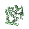
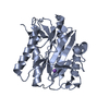
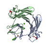
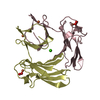
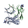
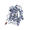
| Structure viewer | Molecule: MolmilJmol/JSmol |
|---|
- Downloads & links
-Download
| PDBx/mmCIF format | 1dfx.cif.gz | 39.1 KB | Display | PDBx/mmCIF format |
|---|---|---|---|---|
| PDB format | pdb1dfx.ent.gz | 27 KB | Display | PDB format |
| PDBx/mmJSON format | 1dfx.json.gz | Tree view | PDBx/mmJSON format | |
| Others |  Other downloads Other downloads |
-Validation report
| Arichive directory | https://data.pdbj.org/pub/pdb/validation_reports/df/1dfxftp://data.pdbj.org/pub/pdb/validation_reports/df/1dfx | HTTPS FTP |
|---|
-Related structure data
| Similar structure data |
|---|


-Links
 PDBj
PDBj- Assembly
| Deposited unit | 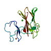
| ||||||||
|---|---|---|---|---|---|---|---|---|---|
| 1 | 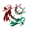
| ||||||||
| Unit cell |
| ||||||||
| Components on special symmetry positions |
|
-Components
| #1: Protein | Mass: 13903.935 Da / Num. of mol.: 1 / Source method: isolated from a natural source / Source: (natural) Desulfovibrio desulfuricans (bacteria) / References: UniProt: P22076 | ||||
|---|---|---|---|---|---|
| #2: Chemical |   Mass: 55.845 Da / Num. of mol.: 2 / Source method: obtained synthetically / Formula: Fe Mass: 55.845 Da / Num. of mol.: 2 / Source method: obtained synthetically / Formula: Fe#3: Chemical | ChemComp-CA / |   Mass: 40.078 Da / Num. of mol.: 1 / Source method: obtained synthetically / Formula: Ca Mass: 40.078 Da / Num. of mol.: 1 / Source method: obtained synthetically / Formula: Ca#4: Water | ChemComp-HOH / |  Mass: 18.015 Da / Num. of mol.: 71 / Source method: isolated from a natural source / Formula: H2O Mass: 18.015 Da / Num. of mol.: 71 / Source method: isolated from a natural source / Formula: H2O |
-Experimental details
-Experiment
| Experiment | Method: X-RAY DIFFRACTION / Number of used crystals: 1 |
|---|
- Sample preparation
| Crystal | Density Matthews: 2.8 Å3/Da / Density % sol: 55 % / Description: MAD DATA COLLECTED AT ESRF BM-14 | ||||||||||||||||||||||||
|---|---|---|---|---|---|---|---|---|---|---|---|---|---|---|---|---|---|---|---|---|---|---|---|---|---|
| Crystal grow | pH: 7.5 Details: PROTEIN WAS CRYSTALLIZED FROM 20% PEG 4000, 0.1 M HEPES, CACL2 0.2 M, PH 7.5 | ||||||||||||||||||||||||
| Crystal | *PLUS | ||||||||||||||||||||||||
| Crystal grow | *PLUS Method: vapor diffusion, sitting drop / Details: Coelho, A.V., (1996) Protein Sci., 5, 1189. | ||||||||||||||||||||||||
| Components of the solutions | *PLUS
|
-Data collection
| Diffraction | Mean temperature: 293 K |
|---|---|
| Diffraction source | Source: SYNCHROTRON / Site: SRS  / Beamline: PX9.5 / Wavelength: 0.994 / Beamline: PX9.5 / Wavelength: 0.994 |
| Detector | Type: MARRESEARCH / Detector: IMAGE PLATE / Date: Oct 1, 1992 / Details: MIRRORS |
| Radiation | Monochromator: SI(111) / Monochromatic (M) / Laue (L): M / Scattering type: x-ray |
| Radiation wavelength | Wavelength: 0.994 Å / Relative weight: 1 |
| Reflection | Resolution: 1.9→20 Å / Num. obs: 11867 / % possible obs: 97.7 % / Observed criterion σ(I): 2 / Redundancy: 2.2 % / Rmerge(I) obs: 0.045 / Rsym value: 0.045 / Net I/σ(I): 8.8 |
| Reflection shell | Resolution: 1.9→1.97 Å / Redundancy: 2.2 % / Rmerge(I) obs: 0.26 / Mean I/σ(I) obs: 2.9 / Rsym value: 0.26 / % possible all: 96.3 |
| Reflection | *PLUS Num. measured all: 25678 |
| Reflection shell | *PLUS % possible obs: 96.3 % |
- Processing
| Software |
| |||||||||||||||||||||||||||||||||
|---|---|---|---|---|---|---|---|---|---|---|---|---|---|---|---|---|---|---|---|---|---|---|---|---|---|---|---|---|---|---|---|---|---|---|
| Refinement | Method to determine structure: MULTIPLE WAVELENGTH ANOMALOUS DISPERSION (MAD) Resolution: 1.9→20 Å / Num. parameters: 4254 / Num. restraintsaints: 3852 / Cross valid method: FREE R / σ(F): 0 / Stereochemistry target values: ENGH & HUBER Details: THE REFINEMENT WAS STARTED WITH X-PLOR. NO GEOMETRICAL RESTRAINTS WERE IMPOSED BETWEEN THE IRON ATOMS AND THEIR LIGANDS.
| |||||||||||||||||||||||||||||||||
| Solvent computation | Solvent model: MOEWS & KRETSINGER | |||||||||||||||||||||||||||||||||
| Refine analyze | Occupancy sum non hydrogen: 1027 | |||||||||||||||||||||||||||||||||
| Refinement step | Cycle: LAST / Resolution: 1.9→20 Å
| |||||||||||||||||||||||||||||||||
| Refine LS restraints |
| |||||||||||||||||||||||||||||||||
| Software | *PLUS Name: SHELXL-93 / Classification: refinement | |||||||||||||||||||||||||||||||||
| Refinement | *PLUS Rfactor all: 0.2 / Rfactor Rwork: 0.2 | |||||||||||||||||||||||||||||||||
| Solvent computation | *PLUS | |||||||||||||||||||||||||||||||||
| Displacement parameters | *PLUS | |||||||||||||||||||||||||||||||||
| Refine LS restraints | *PLUS
|
