[English] 日本語
Yorodumi
- PDB-1vzi: Structure of superoxide reductase bound to ferrocyanide and activ... -

+
Open data


ID or keywords:

Loading...

-
Basic information

Entry
Database: PDB / ID: 1vzi
TitleStructure of superoxide reductase bound to ferrocyanide and active site expansion upon X-ray induced photoreduction
ComponentsDESULFOFERRODOXIN
KeywordsOXIDOREDUCTASE / FERROCYANIDE / MICROSPECTROPHOTOMETRY / REDOX STATES / PHOTOREDUCTION / DINUCLEAR IRON CLUSTER / ELECTRON TRANSPORT
Function / homology
Function and homology information


superoxide reductase / superoxide reductase activity / removal of superoxide radicals / iron ion binding
Similarity search - Function
Desulphoferrodoxin, N-terminal domain / Desulfoferrodoxin / Desulfoferrodoxin, N-terminal domain / Desulfoferrodoxin, N-terminal domain superfamily / Desulfoferrodoxin, N-terminal domain / SOR catalytic domain / Desulfoferrodoxin, ferrous iron-binding domain / Desulfoferrodoxin, ferrous iron-binding domain superfamily / Desulfoferrodoxin / Rubrerythrin, domain 2 ...Desulphoferrodoxin, N-terminal domain / Desulfoferrodoxin / Desulfoferrodoxin, N-terminal domain / Desulfoferrodoxin, N-terminal domain superfamily / Desulfoferrodoxin, N-terminal domain / SOR catalytic domain / Desulfoferrodoxin, ferrous iron-binding domain / Desulfoferrodoxin, ferrous iron-binding domain superfamily / Desulfoferrodoxin / Rubrerythrin, domain 2 / Single Sheet / Immunoglobulin-like / Sandwich / Mainly Beta
Similarity search - Domain/homology
: / Desulfoferrodoxin
Similarity search - Component
Biological speciesDESULFOVIBRIO BAARSII (bacteria)
MethodX-RAY DIFFRACTION / SYNCHROTRON / MOLECULAR REPLACEMENT / Resolution: 1.15 Å
AuthorsAdam, V. / Royant, A. / Niviere, V. / Molina-Heredia, F.P. / Bourgeois, D.
CitationJournal: Structure / Year: 2004
Title: Structure of superoxide reductase bound to ferrocyanide and active site expansion upon X-ray-induced photo-reduction.
Authors: Adam, V. / Royant, A. / Niviere, V. / Molina-Heredia, F.P. / Bourgeois, D.
History
DepositionMay 19, 2004Deposition site: PDBE / Processing site: PDBE
Revision 1.0Aug 27, 2004Provider: repository / Type: Initial release
Revision 1.1Jun 2, 2011Group: Version format compliance
Revision 1.2Jul 13, 2011Group: Version format compliance
Revision 1.3Feb 28, 2018Group: Database references / Source and taxonomy / Category: citation / entity_src_gen
Item: _citation.page_last / _citation.pdbx_database_id_DOI ..._citation.page_last / _citation.pdbx_database_id_DOI / _citation.title / _entity_src_gen.gene_src_common_name / _entity_src_gen.pdbx_gene_src_ncbi_taxonomy_id / _entity_src_gen.pdbx_host_org_ncbi_taxonomy_id / _entity_src_gen.pdbx_host_org_scientific_name / _entity_src_gen.pdbx_host_org_strain
Revision 1.4May 22, 2019Group: Data collection / Refinement description / Category: refine / Item: _refine.pdbx_ls_cross_valid_method
Revision 1.5May 8, 2024Group: Data collection / Database references ...Data collection / Database references / Derived calculations / Other
Category: chem_comp_atom / chem_comp_bond ...chem_comp_atom / chem_comp_bond / database_2 / pdbx_database_status / pdbx_struct_conn_angle / struct_conn
Item: _database_2.pdbx_DOI / _database_2.pdbx_database_accession ..._database_2.pdbx_DOI / _database_2.pdbx_database_accession / _pdbx_database_status.status_code_sf / _pdbx_struct_conn_angle.ptnr1_auth_asym_id / _pdbx_struct_conn_angle.ptnr1_auth_comp_id / _pdbx_struct_conn_angle.ptnr1_auth_seq_id / _pdbx_struct_conn_angle.ptnr1_label_asym_id / _pdbx_struct_conn_angle.ptnr1_label_atom_id / _pdbx_struct_conn_angle.ptnr1_label_comp_id / _pdbx_struct_conn_angle.ptnr1_label_seq_id / _pdbx_struct_conn_angle.ptnr1_symmetry / _pdbx_struct_conn_angle.ptnr2_auth_comp_id / _pdbx_struct_conn_angle.ptnr2_auth_seq_id / _pdbx_struct_conn_angle.ptnr2_label_asym_id / _pdbx_struct_conn_angle.ptnr2_label_atom_id / _pdbx_struct_conn_angle.ptnr2_label_comp_id / _pdbx_struct_conn_angle.ptnr3_auth_asym_id / _pdbx_struct_conn_angle.ptnr3_auth_comp_id / _pdbx_struct_conn_angle.ptnr3_auth_seq_id / _pdbx_struct_conn_angle.ptnr3_label_asym_id / _pdbx_struct_conn_angle.ptnr3_label_atom_id / _pdbx_struct_conn_angle.ptnr3_label_comp_id / _pdbx_struct_conn_angle.ptnr3_label_seq_id / _pdbx_struct_conn_angle.ptnr3_symmetry / _pdbx_struct_conn_angle.value / _struct_conn.pdbx_dist_value / _struct_conn.ptnr1_auth_comp_id / _struct_conn.ptnr1_auth_seq_id / _struct_conn.ptnr1_label_asym_id / _struct_conn.ptnr1_label_atom_id / _struct_conn.ptnr1_label_comp_id / _struct_conn.ptnr1_label_seq_id / _struct_conn.ptnr1_symmetry / _struct_conn.ptnr2_auth_asym_id / _struct_conn.ptnr2_auth_comp_id / _struct_conn.ptnr2_auth_seq_id / _struct_conn.ptnr2_label_asym_id / _struct_conn.ptnr2_label_atom_id / _struct_conn.ptnr2_label_comp_id / _struct_conn.ptnr2_label_seq_id / _struct_conn.ptnr2_symmetry

-
Structure visualization

Structure viewerMolecule:
MolmilJmol/JSmol

Downloads & links

-
Assembly

Deposited unit
A: DESULFOFERRODOXIN
B: DESULFOFERRODOXIN
hetero molecules


Theoretical massNumber of molelcules
Total (without water)28,68212
Polymers28,2372
Non-polymers44510
Water9,332518
1


  • Idetical with deposited unit
  • defined by author&software
TypeNameSymmetry operationNumber
identity operation1_555x,y,z1
MethodPQS
Unit cell
Length a, b, c (Å)123.608, 123.608, 73.016
Angle α, β, γ (deg.)90.00, 90.00, 90.00
Int Tables number98
Space group name H-MI4122
Components on special symmetry positions
IDModelComponents
11A-2063-

HOH

21A-2109-

HOH

31A-2219-

HOH

41B-2121-

HOH

-
Components

#1: Protein DESULFOFERRODOXIN / SUPEROXIDE REDUCTASE / DFX / SOR


Mass: 14118.362 Da / Num. of mol.: 2 / Mutation: YES
Source method: isolated from a genetically manipulated source
Source: (gene. exp.) DESULFOVIBRIO BAARSII (bacteria) / Plasmid: PMLE47A / Production host: Escherichia coli DH5[alpha] (bacteria) / References: UniProt: Q46495, superoxide reductase
#2: Chemical ChemComp-CA / CALCIUM ION


Mass: 40.078 Da / Num. of mol.: 2 / Source method: obtained synthetically / Formula: Ca
#3: Chemical
ChemComp-CL / CHLORIDE ION


Mass: 35.453 Da / Num. of mol.: 4 / Source method: obtained synthetically / Formula: Cl
#4: Chemical
ChemComp-FE2 / FE (II) ION


Mass: 55.845 Da / Num. of mol.: 4 / Source method: obtained synthetically / Formula: Fe
#5: Water ChemComp-HOH / water


Mass: 18.015 Da / Num. of mol.: 518 / Source method: isolated from a natural source / Formula: H2O
Compound details
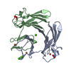
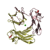
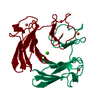
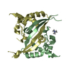
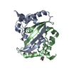
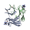
NON-HEME IRON PROTEIN. CATALYSES SUPEROXIDE ANION + 2 H(+) = H(2)O(2). COFACTOR: BINDS 1 ...NON-HEME IRON PROTEIN. CATALYSES SUPEROXIDE ANION + 2 H(+) = H(2)O(2). COFACTOR: BINDS 1 DESULFOFERRODOXIN-LIKE FES(4) SITE AND 1 ENGINEERED MUTATION GLU 47 TO ALA, CHAINS A AND B

-
Experimental details

-
Experiment

ExperimentMethod: X-RAY DIFFRACTION / Number of used crystals: 1

-
Sample preparation

CrystalDensity Matthews: 2.47 Å3/Da / Density % sol: 38 %
Crystal growpH: 8 / Details: 18% PEG 4000, 100 MM TRIS PH 8.0, 100 MM CACL2

-
Data collection

DiffractionMean temperature: 100 K
Diffraction sourceSource: SYNCHROTRON / Site: ESRF / Beamline: ID14-2 / Wavelength: 0.923
DetectorType: ADSC CCD / Detector: CCD / Date: Jul 23, 2003 / Details: MIRRORS
RadiationMonochromator: DIAMOND 111 / Protocol: SINGLE WAVELENGTH / Monochromatic (M) / Laue (L): M / Scattering type: x-ray
Radiation wavelengthWavelength: 0.923 Å / Relative weight: 1
ReflectionResolution: 1.15→45 Å / Num. obs: 93621 / % possible obs: 99 % / Redundancy: 6.1 % / Rmerge(I) obs: 0.05 / Net I/σ(I): 13.6
Reflection shellRmerge(I) obs: 0.41 / % possible all: 98.1

-
Processing

Software
NameClassification
SHELXL-97refinement
XDSdata reduction
XDSdata scaling
CNSphasing
RefinementMethod to determine structure: MOLECULAR REPLACEMENT / Resolution: 1.15→45 Å / Num. parameters: 22352 / Num. restraintsaints: 0 / Cross valid method: FREE R-VALUE / σ(F): 0 / Stereochemistry target values: ENGH AND HUBER
Details: ANISOTROPIC REFINEMENT REDUCED FREE R (NO CUTOFF). CHLORIDE ION IN ACTIVE SITE II (MONOMER B) IS MODELED IN DUAL CONFORMATIONS
RfactorNum. reflection% reflectionSelection details
all0.1427 98585 --
obs--99.1 %-
Rfree---RANDOM
Refine analyzeNum. disordered residues: 1 / Occupancy sum hydrogen: 0 / Occupancy sum non hydrogen: 2480.25
Refinement stepCycle: LAST / Resolution: 1.15→45 Å
ProteinNucleic acidLigandSolventTotal
Num. atoms1956 0 10 518 2484
Refine LS restraints
Refine-IDTypeDev ideal
X-RAY DIFFRACTIONs_bond_d0.016
X-RAY DIFFRACTIONs_angle_d0.034
X-RAY DIFFRACTIONs_similar_dist
X-RAY DIFFRACTIONs_from_restr_planes
X-RAY DIFFRACTIONs_zero_chiral_vol
X-RAY DIFFRACTIONs_non_zero_chiral_vol
X-RAY DIFFRACTIONs_anti_bump_dis_restr
X-RAY DIFFRACTIONs_rigid_bond_adp_cmpnt
X-RAY DIFFRACTIONs_similar_adp_cmpnt
X-RAY DIFFRACTIONs_approx_iso_adps

+
About Yorodumi

-
News

-
Feb 9, 2022. New format data for meta-information of EMDB entries

New format data for meta-information of EMDB entries

  • Version 3 of the EMDB header file is now the official format.
  • The previous official version 1.9 will be removed from the archive.

Related info.:EMDB header

External links:wwPDB to switch to version 3 of the EMDB data model

-
Aug 12, 2020. Covid-19 info

Covid-19 info

URL: https://pdbjlvh1.pdbj.org/emnavi/covid19.php

New page: Covid-19 featured information page in EM Navigator.

Related info.:Covid-19 info / Mar 5, 2020. Novel coronavirus structure data

+
Mar 5, 2020. Novel coronavirus structure data

Novel coronavirus structure data

Related info.:Yorodumi Speices / Aug 12, 2020. Covid-19 info

External links:COVID-19 featured content - PDBj / Molecule of the Month (242):Coronavirus Proteases

+
Jan 31, 2019. EMDB accession codes are about to change! (news from PDBe EMDB page)

EMDB accession codes are about to change! (news from PDBe EMDB page)

  • The allocation of 4 digits for EMDB accession codes will soon come to an end. Whilst these codes will remain in use, new EMDB accession codes will include an additional digit and will expand incrementally as the available range of codes is exhausted. The current 4-digit format prefixed with “EMD-” (i.e. EMD-XXXX) will advance to a 5-digit format (i.e. EMD-XXXXX), and so on. It is currently estimated that the 4-digit codes will be depleted around Spring 2019, at which point the 5-digit format will come into force.
  • The EM Navigator/Yorodumi systems omit the EMD- prefix.

Related info.:Q: What is EMD? / ID/Accession-code notation in Yorodumi/EM Navigator

External links:EMDB Accession Codes are Changing Soon! / Contact to PDBj

+
Jul 12, 2017. Major update of PDB

Major update of PDB

  • wwPDB released updated PDB data conforming to the new PDBx/mmCIF dictionary.
  • This is a major update changing the version number from 4 to 5, and with Remediation, in which all the entries are updated.
  • In this update, many items about electron microscopy experimental information are reorganized (e.g. em_software).
  • Now, EM Navigator and Yorodumi are based on the updated data.

External links:wwPDB Remediation / Enriched Model Files Conforming to OneDep Data Standards Now Available in the PDB FTP Archive

-
Yorodumi

Thousand views of thousand structures

  • Yorodumi is a browser for structure data from EMDB, PDB, SASBDB, etc.
  • This page is also the successor to EM Navigator detail page, and also detail information page/front-end page for Omokage search.
  • The word "yorodu" (or yorozu) is an old Japanese word meaning "ten thousand". "mi" (miru) is to see.

Related info.:EMDB / PDB / SASBDB / Comparison of 3 databanks / Yorodumi Search / Aug 31, 2016. New EM Navigator & Yorodumi / Yorodumi Papers / Jmol/JSmol / Function and homology information / Changes in new EM Navigator and Yorodumi

Read more