[English] 日本語
Yorodumi
- PDB-1vzg: Structure of superoxide reductase bound to ferrocyanide and activ... -

+
Open data


ID or keywords:

Loading...

-
Basic information

Entry
Database: PDB / ID: 1vzg
TitleStructure of superoxide reductase bound to ferrocyanide and active site expansion upon X-ray induced photoreduction
ComponentsDESULFOFERRODOXIN
KeywordsOXIDOREDUCTASE / FERROCYANIDE / MICROSPECTROPHOTOMETRY / REDOX STATES / PHOTOREDUCTION / DINUCLEAR IRON CLUSTER / ELECTRON TRANSPORT
Function / homology
Function and homology information


superoxide reductase / superoxide reductase activity / removal of superoxide radicals / iron ion binding
Similarity search - Function
Desulphoferrodoxin, N-terminal domain / Desulfoferrodoxin / Desulfoferrodoxin, N-terminal domain / Desulfoferrodoxin, N-terminal domain superfamily / Desulfoferrodoxin, N-terminal domain / SOR catalytic domain / Desulfoferrodoxin, ferrous iron-binding domain / Desulfoferrodoxin, ferrous iron-binding domain superfamily / Desulfoferrodoxin / Rubrerythrin, domain 2 ...Desulphoferrodoxin, N-terminal domain / Desulfoferrodoxin / Desulfoferrodoxin, N-terminal domain / Desulfoferrodoxin, N-terminal domain superfamily / Desulfoferrodoxin, N-terminal domain / SOR catalytic domain / Desulfoferrodoxin, ferrous iron-binding domain / Desulfoferrodoxin, ferrous iron-binding domain superfamily / Desulfoferrodoxin / Rubrerythrin, domain 2 / Single Sheet / Immunoglobulin-like / Sandwich / Mainly Beta
Similarity search - Domain/homology
HEXACYANOFERRATE(3-) / : / Desulfoferrodoxin
Similarity search - Component
Biological speciesDESULFOVIBRIO BAARSII (bacteria)
MethodX-RAY DIFFRACTION / SYNCHROTRON / MOLECULAR REPLACEMENT / Resolution: 1.69 Å
AuthorsAdam, V. / Royant, A. / Niviere, V. / Molina-Heredia, F.P. / Bourgeois, D.
CitationJournal: Structure / Year: 2004
Title: Structure of Superoxide Reductase Bound to Ferrocyanide and Active Site Expansion Upon X-Ray Induced Photoreduction
Authors: Adam, V. / Royant, A. / Niviere, V. / Molina-Heredia, F.P. / Bourgeois, D.
History
DepositionMay 19, 2004Deposition site: PDBE / Processing site: PDBE
Revision 1.0Aug 27, 2004Provider: repository / Type: Initial release
Revision 1.1Jun 2, 2011Group: Version format compliance
Revision 1.2Jul 13, 2011Group: Version format compliance
Revision 1.3Dec 13, 2023Group: Data collection / Database references ...Data collection / Database references / Derived calculations / Other / Refinement description
Category: chem_comp_atom / chem_comp_bond ...chem_comp_atom / chem_comp_bond / database_2 / pdbx_database_status / pdbx_initial_refinement_model / pdbx_struct_conn_angle / struct_conn
Item: _database_2.pdbx_DOI / _database_2.pdbx_database_accession ..._database_2.pdbx_DOI / _database_2.pdbx_database_accession / _pdbx_database_status.status_code_sf / _pdbx_struct_conn_angle.ptnr1_auth_asym_id / _pdbx_struct_conn_angle.ptnr1_auth_comp_id / _pdbx_struct_conn_angle.ptnr1_auth_seq_id / _pdbx_struct_conn_angle.ptnr1_label_asym_id / _pdbx_struct_conn_angle.ptnr1_label_atom_id / _pdbx_struct_conn_angle.ptnr1_label_comp_id / _pdbx_struct_conn_angle.ptnr1_label_seq_id / _pdbx_struct_conn_angle.ptnr2_auth_comp_id / _pdbx_struct_conn_angle.ptnr2_auth_seq_id / _pdbx_struct_conn_angle.ptnr2_label_asym_id / _pdbx_struct_conn_angle.ptnr2_label_atom_id / _pdbx_struct_conn_angle.ptnr2_label_comp_id / _pdbx_struct_conn_angle.ptnr3_auth_asym_id / _pdbx_struct_conn_angle.ptnr3_auth_comp_id / _pdbx_struct_conn_angle.ptnr3_auth_seq_id / _pdbx_struct_conn_angle.ptnr3_label_asym_id / _pdbx_struct_conn_angle.ptnr3_label_atom_id / _pdbx_struct_conn_angle.ptnr3_label_comp_id / _pdbx_struct_conn_angle.ptnr3_label_seq_id / _pdbx_struct_conn_angle.value / _struct_conn.pdbx_dist_value / _struct_conn.pdbx_ptnr1_label_alt_id / _struct_conn.pdbx_ptnr2_label_alt_id / _struct_conn.ptnr1_auth_comp_id / _struct_conn.ptnr1_auth_seq_id / _struct_conn.ptnr1_label_asym_id / _struct_conn.ptnr1_label_atom_id / _struct_conn.ptnr1_label_comp_id / _struct_conn.ptnr1_label_seq_id / _struct_conn.ptnr2_auth_asym_id / _struct_conn.ptnr2_auth_comp_id / _struct_conn.ptnr2_auth_seq_id / _struct_conn.ptnr2_label_asym_id / _struct_conn.ptnr2_label_atom_id / _struct_conn.ptnr2_label_comp_id / _struct_conn.ptnr2_label_seq_id

-
Structure visualization

Structure viewerMolecule:
MolmilJmol/JSmol

Downloads & links

-
Assembly

Deposited unit
A: DESULFOFERRODOXIN
B: DESULFOFERRODOXIN
hetero molecules


Theoretical massNumber of molelcules
Total (without water)28,9249
Polymers28,2372
Non-polymers6877
Water2,900161
1


  • Idetical with deposited unit
  • defined by author&software
TypeNameSymmetry operationNumber
identity operation1_555x,y,z1
MethodPQS
Unit cell
Length a, b, c (Å)42.130, 67.650, 82.190
Angle α, β, γ (deg.)90.00, 90.00, 90.00
Int Tables number19
Space group name H-MP212121

-
Components

#1: Protein DESULFOFERRODOXIN / SUPEROXIDE REDUCTASE / DFX / SOR


Mass: 14118.362 Da / Num. of mol.: 2 / Mutation: YES
Source method: isolated from a genetically manipulated source
Source: (gene. exp.) DESULFOVIBRIO BAARSII (bacteria) / Plasmid: PMLE47A / Production host: ESCHERICHIA COLI (E. coli) / Strain (production host): DH5 ALPHA / References: UniProt: Q46495, superoxide reductase
#2: Chemical
ChemComp-FE / FE (III) ION


Mass: 55.845 Da / Num. of mol.: 4 / Source method: obtained synthetically / Formula: Fe
#3: Chemical ChemComp-CA / CALCIUM ION


Mass: 40.078 Da / Num. of mol.: 1 / Source method: obtained synthetically / Formula: Ca
#4: Chemical ChemComp-FC6 / HEXACYANOFERRATE(3-) / FERRI(III)HEXACYANIDE


Mass: 211.949 Da / Num. of mol.: 2 / Source method: obtained synthetically / Formula: C6FeN6
#5: Water ChemComp-HOH / water


Mass: 18.015 Da / Num. of mol.: 161 / Source method: isolated from a natural source / Formula: H2O
Compound details
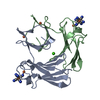
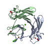
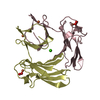
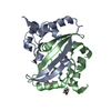
NON-HEME IRON PROTEIN. CATALYSES SUPEROXIDE ANION + 2 H(+) = H(2)O(2). COFACTOR: BINDS 1 ...NON-HEME IRON PROTEIN. CATALYSES SUPEROXIDE ANION + 2 H(+) = H(2)O(2). COFACTOR: BINDS 1 DESULFOFERRODOXIN-LIKE FES(4) SITE AND 1 ENGINEERED MUTATION GLU 47 TO ALA, CHAINS A AND B

-
Experimental details

-
Experiment

ExperimentMethod: X-RAY DIFFRACTION / Number of used crystals: 1

-
Sample preparation

CrystalDensity Matthews: 2.07 Å3/Da / Density % sol: 36 %
Crystal growpH: 8 / Details: 18% PEG 4000, 100 MM TRIS PH 8.0, 100 MM CACL2

-
Data collection

DiffractionMean temperature: 100 K
Diffraction sourceSource: SYNCHROTRON / Site: ESRF / Beamline: ID14-4 / Wavelength: 0.92
DetectorType: ADSC CCD / Detector: CCD / Date: Jun 29, 2003 / Details: MIRRORS
RadiationMonochromator: SI 111 / Monochromatic (M) / Laue (L): M / Scattering type: x-ray
Radiation wavelengthWavelength: 0.92 Å / Relative weight: 1
ReflectionResolution: 1.69→37.49 Å / Num. obs: 26217 / % possible obs: 98.3 % / Observed criterion σ(I): 2 / Redundancy: 2.9 % / Biso Wilson estimate: 17.6 Å2 / Rmerge(I) obs: 0.08 / Net I/σ(I): 12.3
Reflection shellRedundancy: 2.9 % / Rmerge(I) obs: 0.32 / Mean I/σ(I) obs: 3.7 / % possible all: 94.1

-
Processing

Software
NameVersionClassification
CNS1refinement
MOSFLMdata reduction
SCALAdata scaling
CNSphasing
RefinementMethod to determine structure: MOLECULAR REPLACEMENT
Starting model: PDB ENTRY 1DFX
Resolution: 1.69→37.49 Å / Rfactor Rfree error: 0.007 / Data cutoff high absF: 1920830.55 / Isotropic thermal model: RESTRAINED / Cross valid method: THROUGHOUT / σ(F): 0
Details: CYSTEINES FROM THE CENTER I WERE MODELED IN ALTERNATE CONFORMATIONS, AS WELL AS CENTER I IRONS AND RESIDUES 84 TO 86
RfactorNum. reflection% reflectionSelection details
Rfree0.249 1285 5 %RANDOM
Rwork0.205 ---
obs0.205 25836 96.4 %-
Solvent computationSolvent model: FLAT MODEL / Bsol: 40.1442 Å2 / ksol: 0.373404 e/Å3
Displacement parametersBiso mean: 18.4 Å2
Baniso -1Baniso -2Baniso -3
1-0.11 Å20 Å20 Å2
2--0.1 Å20 Å2
3----0.21 Å2
Refine analyze
FreeObs
Luzzati coordinate error0.24 Å0.2 Å
Luzzati d res low-5 Å
Luzzati sigma a0.13 Å0.12 Å
Refinement stepCycle: LAST / Resolution: 1.69→37.49 Å
ProteinNucleic acidLigandSolventTotal
Num. atoms1956 0 31 161 2148
Refine LS restraints
Refine-IDTypeDev idealDev ideal target
X-RAY DIFFRACTIONc_bond_d0.005
X-RAY DIFFRACTIONc_bond_d_na
X-RAY DIFFRACTIONc_bond_d_prot
X-RAY DIFFRACTIONc_angle_d
X-RAY DIFFRACTIONc_angle_d_na
X-RAY DIFFRACTIONc_angle_d_prot
X-RAY DIFFRACTIONc_angle_deg1.4
X-RAY DIFFRACTIONc_angle_deg_na
X-RAY DIFFRACTIONc_angle_deg_prot
X-RAY DIFFRACTIONc_dihedral_angle_d26.4
X-RAY DIFFRACTIONc_dihedral_angle_d_na
X-RAY DIFFRACTIONc_dihedral_angle_d_prot
X-RAY DIFFRACTIONc_improper_angle_d4.77
X-RAY DIFFRACTIONc_improper_angle_d_na
X-RAY DIFFRACTIONc_improper_angle_d_prot
X-RAY DIFFRACTIONc_mcbond_it1.261.5
X-RAY DIFFRACTIONc_mcangle_it1.962
X-RAY DIFFRACTIONc_scbond_it1.982
X-RAY DIFFRACTIONc_scangle_it2.892.5
LS refinement shellResolution: 1.69→1.8 Å / Rfactor Rfree error: 0.022 / Total num. of bins used: 6
RfactorNum. reflection% reflection
Rfree0.316 209 5.2 %
Rwork0.262 --
obs--91.9 %
Xplor file
Refine-IDSerial noParam fileTopol file
X-RAY DIFFRACTION1PROTEIN_REP.PARAMPROTEIN.TOP
X-RAY DIFFRACTION2WATER_REP.PARAMWATER.TOP
X-RAY DIFFRACTION3ION.PARAMION.TOP
X-RAY DIFFRACTION4FER.PARAMFER.TOP
X-RAY DIFFRACTION5PROTEIN-CIS.PARAM

+
About Yorodumi

-
News

-
Feb 9, 2022. New format data for meta-information of EMDB entries

New format data for meta-information of EMDB entries

  • Version 3 of the EMDB header file is now the official format.
  • The previous official version 1.9 will be removed from the archive.

Related info.:EMDB header

External links:wwPDB to switch to version 3 of the EMDB data model

-
Aug 12, 2020. Covid-19 info

Covid-19 info

URL: https://pdbjlvh1.pdbj.org/emnavi/covid19.php

New page: Covid-19 featured information page in EM Navigator.

Related info.:Covid-19 info / Mar 5, 2020. Novel coronavirus structure data

+
Mar 5, 2020. Novel coronavirus structure data

Novel coronavirus structure data

Related info.:Yorodumi Speices / Aug 12, 2020. Covid-19 info

External links:COVID-19 featured content - PDBj / Molecule of the Month (242):Coronavirus Proteases

+
Jan 31, 2019. EMDB accession codes are about to change! (news from PDBe EMDB page)

EMDB accession codes are about to change! (news from PDBe EMDB page)

  • The allocation of 4 digits for EMDB accession codes will soon come to an end. Whilst these codes will remain in use, new EMDB accession codes will include an additional digit and will expand incrementally as the available range of codes is exhausted. The current 4-digit format prefixed with “EMD-” (i.e. EMD-XXXX) will advance to a 5-digit format (i.e. EMD-XXXXX), and so on. It is currently estimated that the 4-digit codes will be depleted around Spring 2019, at which point the 5-digit format will come into force.
  • The EM Navigator/Yorodumi systems omit the EMD- prefix.

Related info.:Q: What is EMD? / ID/Accession-code notation in Yorodumi/EM Navigator

External links:EMDB Accession Codes are Changing Soon! / Contact to PDBj

+
Jul 12, 2017. Major update of PDB

Major update of PDB

  • wwPDB released updated PDB data conforming to the new PDBx/mmCIF dictionary.
  • This is a major update changing the version number from 4 to 5, and with Remediation, in which all the entries are updated.
  • In this update, many items about electron microscopy experimental information are reorganized (e.g. em_software).
  • Now, EM Navigator and Yorodumi are based on the updated data.

External links:wwPDB Remediation / Enriched Model Files Conforming to OneDep Data Standards Now Available in the PDB FTP Archive

-
Yorodumi

Thousand views of thousand structures

  • Yorodumi is a browser for structure data from EMDB, PDB, SASBDB, etc.
  • This page is also the successor to EM Navigator detail page, and also detail information page/front-end page for Omokage search.
  • The word "yorodu" (or yorozu) is an old Japanese word meaning "ten thousand". "mi" (miru) is to see.

Related info.:EMDB / PDB / SASBDB / Comparison of 3 databanks / Yorodumi Search / Aug 31, 2016. New EM Navigator & Yorodumi / Yorodumi Papers / Jmol/JSmol / Function and homology information / Changes in new EM Navigator and Yorodumi

Read more