 Movie
Movie Controller

[English] 日本語
 Yorodumi
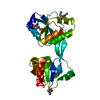
- PDB-1deg: THE LINKER OF DES-GLU84 CALMODULIN IS BENT AS SEEN IN THE CRYSTAL... -
+ Open data

- Basic information
| Entry | Database: PDB / ID: 1deg | ||||||
|---|---|---|---|---|---|---|---|
| Title | THE LINKER OF DES-GLU84 CALMODULIN IS BENT AS SEEN IN THE CRYSTAL STRUCTURE | ||||||
 Components Components | CALMODULIN | ||||||
 Keywords Keywords | CALCIUM-BINDING PROTEIN | ||||||
| Function / homology |  Function and homology information Function and homology informationregulation of store-operated calcium channel activity / : / : / regulation of response to tumor cell / positive regulation of autophagic cell death / DAPK1-calmodulin complex / : / : / : / : ...regulation of store-operated calcium channel activity / : / : / regulation of response to tumor cell / positive regulation of autophagic cell death / DAPK1-calmodulin complex / : / : / : / : / : / : / type 3 metabotropic glutamate receptor binding / establishment of protein localization to membrane / positive regulation of DNA binding / negative regulation of high voltage-gated calcium channel activity / negative regulation of ryanodine-sensitive calcium-release channel activity / organelle localization by membrane tethering / : / autophagosome membrane docking / regulation of synaptic vesicle exocytosis / negative regulation of calcium ion export across plasma membrane / regulation of ryanodine-sensitive calcium-release channel activity / regulation of cardiac muscle cell action potential / calcineurin-mediated signaling / nitric-oxide synthase binding / adenylate cyclase binding / protein phosphatase activator activity / regulation of synaptic vesicle endocytosis / detection of calcium ion / regulation of cardiac muscle contraction / catalytic complex / positive regulation of nitric-oxide synthase activity / phosphatidylinositol 3-kinase binding / activation of adenylate cyclase activity / calcium channel inhibitor activity / regulation of release of sequestered calcium ion into cytosol by sarcoplasmic reticulum / enzyme regulator activity / titin binding / regulation of calcium-mediated signaling / voltage-gated potassium channel complex / calcium channel complex / potassium ion transmembrane transport / regulation of heart rate / nitric-oxide synthase regulator activity / adenylate cyclase activator activity / regulation of cytokinesis / spindle microtubule / response to amphetamine / sarcomere / calcium channel regulator activity / calcium-mediated signaling / response to calcium ion / G2/M transition of mitotic cell cycle / spindle pole / disordered domain specific binding / calcium-dependent protein binding / protein autophosphorylation / myelin sheath / synaptic vesicle membrane / growth cone / vesicle / transmembrane transporter binding / neuron projection / positive regulation of apoptotic process / protein domain specific binding / calcium ion binding / centrosome / protein kinase binding / protein-containing complex / mitochondrion / nucleoplasm / nucleus / plasma membrane / cytoplasm / cytosol Similarity search - Function | ||||||
| Biological species |  | ||||||
| Method |  X-RAY DIFFRACTION / Resolution: 2.9 Å X-RAY DIFFRACTION / Resolution: 2.9 Å | ||||||
 Authors Authors | Raghunathan, S. / Chandross, R. / Cheng, B.P. / Persechini, A. / Sobottk, S.E. / Kretsinger, R.H. | ||||||
 Citation Citation | Journal: Proc.Natl.Acad.Sci.Usa / Year: 1993 Title: The linker of des-Glu84-calmodulin is bent. Authors: Raghunathan, S. / Chandross, R.J. / Cheng, B.P. / Persechini, A. / Sobottka, S.E. / Kretsinger, R.H. #1: Journal: Biochemistry / Year: 1991Title: Small-Angle X-Ray Scattering Studies of Calmodulin Mutants with Deletions in the Linker Region of the Central Helix Indicate that the Linker Region Retains a Predominantly A-Helical Conformation Authors: Kataoka, M. / Head, J.F. / Persechini, A. / Kretsinger, R.H. / Engelman, D.M. #2: Journal: Proc.Natl.Acad.Sci.USA / Year: 1991Title: Calmodulins with Deletions in the Central Helix Functionally Replace the Native Protein in Yeast Cells Authors: Persechini, A. / Kretsinger, R.H. / Davis, T.N. #3: Journal: J.Biol.Chem. / Year: 1989Title: The Effects of Deletions in the Central Helix of Calmodulin on Enzyme Activation and Peptide Binding Authors: Persechini, A. / Blumenthal, D.K. / Jarrett, H.W. / Klee, C.B. / Hardy, D.O. / Kretsinger, R.H. #4: Journal: J.Biol.Chem. / Year: 1988Title: The Central Helix of Calmodulin Functions as a Flexible Tether Authors: Persechini, A. / Kretsinger, R.H. #5: Journal: J.CARDIOVASC.PHARMACOL. / Year: 1988Title: Toward a Model of the Calmodulin-Myosin Light Chain Kinase Complex: Implications of Calmodulin Function Authors: Persechini, A. / Kretsinger, R.H. | ||||||
| History |
| ||||||
| Remark 650 | HELIX THERE IS A BEND OF 40 DEGREES BETWEEN THE F2 HELIX RESIDUES 66 - 76, AND THE LINKER 77 - 83. ...HELIX THERE IS A BEND OF 40 DEGREES BETWEEN THE F2 HELIX RESIDUES 66 - 76, AND THE LINKER 77 - 83. THE ANGLE BETWEEN THE LINKER AND THE E3 HELIX, 85 - 92, IS 85 DEGREES. |
- Structure visualization
| Structure viewer | Molecule: MolmilJmol/JSmol |
|---|
- Downloads & links
-Download
| PDBx/mmCIF format | 1deg.cif.gz | 16.8 KB | Display | PDBx/mmCIF format |
|---|---|---|---|---|
| PDB format | pdb1deg.ent.gz | 7.5 KB | Display | PDB format |
| PDBx/mmJSON format | 1deg.json.gz | Tree view | PDBx/mmJSON format | |
| Others |  Other downloads Other downloads |
-Validation report
| Arichive directory | https://data.pdbj.org/pub/pdb/validation_reports/de/1degftp://data.pdbj.org/pub/pdb/validation_reports/de/1deg | HTTPS FTP |
|---|
-Related structure data
| Similar structure data |
|---|


-Links
 PDBj
PDBj

- Assembly
| Deposited unit | 
| ||||||||
|---|---|---|---|---|---|---|---|---|---|
| 1 |
| ||||||||
| Unit cell |
| ||||||||
| Noncrystallographic symmetry (NCS) | NCS oper: (Code: given Matrix: (-0.990874, 0.076905, 0.110695), Vector: Details | THERE IS AN APPROXIMATE NON-CRYSTALLOGRAPHIC TWO-FOLD AXIS ROUGHLY PARALLEL TO B, RELATING DOMAIN I, (RESIDUES 12 - 74) AND DOMAIN II (RESIDUES 85 - 147). THE TRANSFORMATION PRESENTED ON *MTRIX* RECORDS BELOW WILL YIELD APPROXIMATE COORDINATES FOR DOMAIN I WHEN APPLIED TO DOMAIN II. | |
-Components
| #1: Protein | Mass: 16034.614 Da / Num. of mol.: 1 Source method: isolated from a genetically manipulated source Source: (gene. exp.) | ||
|---|---|---|---|
| #2: Chemical | ChemComp-CA /   Mass: 40.078 Da / Num. of mol.: 4 / Source method: obtained synthetically / Formula: Ca Mass: 40.078 Da / Num. of mol.: 4 / Source method: obtained synthetically / Formula: CaSequence details | SEQUENCE ADVISORY NOTICE DIFFERENCE BETWEEN SWISS-PROT AND PDB SEQUENCE. SWISS-PROT ENTRY NAME: ...SEQUENCE ADVISORY NOTICE DIFFERENCE | |
-Experimental details
-Experiment
| Experiment | Method: X-RAY DIFFRACTION |
|---|
- Sample preparation
| Crystal | Density Matthews: 2.2 Å3/Da / Density % sol: 44.06 % | ||||||||||||||||||||||||||||||
|---|---|---|---|---|---|---|---|---|---|---|---|---|---|---|---|---|---|---|---|---|---|---|---|---|---|---|---|---|---|---|---|
| Crystal grow | *PLUS Temperature: 4 ℃ / pH: 6.1 / Method: vapor diffusion | ||||||||||||||||||||||||||||||
| Components of the solutions | *PLUS
|
-Data collection
| Radiation | Scattering type: x-ray |
|---|---|
| Radiation wavelength | Relative weight: 1 |
| Reflection | *PLUS Highest resolution: 2.9 Å / Num. all: 3483 / Num. obs: 3396 / Observed criterion σ(I): 0 / Redundancy: 3.8 % / Rmerge(I) obs: 0.09 |
| Reflection shell | *PLUS Highest resolution: 2.9 Å / Lowest resolution: 3.1 Å / Redundancy: 2.1 % / Rmerge(I) obs: 0.115 / Mean I/σ(I) obs: 5.3 |
- Processing
| Software |
| ||||||||||||||||||||||||||||||||||||||||||||||||||||||||||||
|---|---|---|---|---|---|---|---|---|---|---|---|---|---|---|---|---|---|---|---|---|---|---|---|---|---|---|---|---|---|---|---|---|---|---|---|---|---|---|---|---|---|---|---|---|---|---|---|---|---|---|---|---|---|---|---|---|---|---|---|---|---|
| Refinement | Rfactor Rwork: 0.23 / Rfactor obs: 0.23 / Highest resolution: 2.9 Å | ||||||||||||||||||||||||||||||||||||||||||||||||||||||||||||
| Refinement step | Cycle: LAST / Highest resolution: 2.9 Å
| ||||||||||||||||||||||||||||||||||||||||||||||||||||||||||||
| Refine LS restraints |
|
