 Movie
Movie Controller

[English] 日本語
 Yorodumi
- PDB-2a7l: Structure of the human hypothetical ubiquitin-conjugating enzyme,... -
+ Open data

- Basic information
| Entry | Database: PDB / ID: 2a7l | ||||||
|---|---|---|---|---|---|---|---|
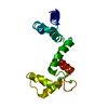
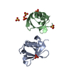
| Title | Structure of the human hypothetical ubiquitin-conjugating enzyme, LOC55284 | ||||||
 Components Components | Hypothetical ubiquitin-conjugating enzyme LOC55284 | ||||||
 Keywords Keywords | LIGASE / STRUCTURAL GENOMICS CONSORTIUM / (SGC) / UBIQUITIN / UBIQUITIN- CONJUGATING ENZYME | ||||||
| Function / homology |  Function and homology information Function and homology informationN-terminal E2 ubiquitin-conjugating enzyme / protein K11-linked ubiquitination / cellular response to misfolded protein / E2 ubiquitin-conjugating enzyme / ubiquitin conjugating enzyme activity / protein quality control for misfolded or incompletely synthesized proteins / protein monoubiquitination / negative regulation of TORC1 signaling / antiviral innate immune response / Synthesis of active ubiquitin: roles of E1 and E2 enzymes ...N-terminal E2 ubiquitin-conjugating enzyme / protein K11-linked ubiquitination / cellular response to misfolded protein / E2 ubiquitin-conjugating enzyme / ubiquitin conjugating enzyme activity / protein quality control for misfolded or incompletely synthesized proteins / protein monoubiquitination / negative regulation of TORC1 signaling / antiviral innate immune response / Synthesis of active ubiquitin: roles of E1 and E2 enzymes / protein polyubiquitination / ubiquitin-protein transferase activity / Antigen processing: Ubiquitination & Proteasome degradation / proteasome-mediated ubiquitin-dependent protein catabolic process / DNA repair / ubiquitin protein ligase binding / nucleoplasm / ATP binding / nucleus / cytoplasm Similarity search - Function | ||||||
| Biological species |  Homo sapiens (human) Homo sapiens (human) | ||||||
| Method |  X-RAY DIFFRACTION / MOLECULAR REPLACEMENT / Resolution: 1.82 Å X-RAY DIFFRACTION / MOLECULAR REPLACEMENT / Resolution: 1.82 Å | ||||||
 Authors Authors | Walker, J.R. / Avvakumov, G.V. / Xue, S. / Newman, E.M. / Mackenzie, F. / Weigelt, J. / Sundstrom, M. / Arrowsmith, C. / Edwards, A. / Bochkarev, A. ...Walker, J.R. / Avvakumov, G.V. / Xue, S. / Newman, E.M. / Mackenzie, F. / Weigelt, J. / Sundstrom, M. / Arrowsmith, C. / Edwards, A. / Bochkarev, A. / Dhe-Paganon, S. / Structural Genomics Consortium (SGC) | ||||||
 Citation Citation | Journal: Mol Cell Proteomics / Year: 2012 Title: A human ubiquitin conjugating enzyme (E2)-HECT E3 ligase structure-function screen. Authors: Sheng, Y. / Hong, J.H. / Doherty, R. / Srikumar, T. / Shloush, J. / Avvakumov, G.V. / Walker, J.R. / Xue, S. / Neculai, D. / Wan, J.W. / Kim, S.K. / Arrowsmith, C.H. / Raught, B. / Dhe-Paganon, S. | ||||||
| History |
|
- Structure visualization
| Structure viewer | Molecule: MolmilJmol/JSmol |
|---|
- Downloads & links
-Download
| PDBx/mmCIF format | 2a7l.cif.gz | 67 KB | Display | PDBx/mmCIF format |
|---|---|---|---|---|
| PDB format | pdb2a7l.ent.gz | 48.7 KB | Display | PDB format |
| PDBx/mmJSON format | 2a7l.json.gz | Tree view | PDBx/mmJSON format | |
| Others |  Other downloads Other downloads |
-Validation report
| Arichive directory | https://data.pdbj.org/pub/pdb/validation_reports/a7/2a7lftp://data.pdbj.org/pub/pdb/validation_reports/a7/2a7l | HTTPS FTP |
|---|
-Related structure data
| Related structure data |  1y6lSC  1yh2C  1yrvC  1zdnC  1zuoC  2a4dC  2awfC  2f4wC  2ob4C  2qgxC  2z5dC  3bzhC  3cegC S: Starting model for refinement C: citing same article ( |
|---|---|
| Similar structure data |


-Links
 PDBj
PDBj

- Assembly
| Deposited unit | 
| ||||||||
|---|---|---|---|---|---|---|---|---|---|
| 1 | 
| ||||||||
| 2 | 
| ||||||||
| Unit cell |
| ||||||||
| Details | The biological unit is a monomer. |
-Components
| #1: Protein | Mass: 15159.265 Da / Num. of mol.: 2 Source method: isolated from a genetically manipulated source Source: (gene. exp.) Homo sapiens (human) / Gene: FLJ11011 / Plasmid: PET28-LIC / Species (production host): Escherichia coli / Production host:  #2: Chemical |   Mass: 22.990 Da / Num. of mol.: 2 / Source method: obtained synthetically / Formula: Na Mass: 22.990 Da / Num. of mol.: 2 / Source method: obtained synthetically / Formula: Na#3: Water | ChemComp-HOH / |  Mass: 18.015 Da / Num. of mol.: 200 / Source method: isolated from a natural source / Formula: H2O Mass: 18.015 Da / Num. of mol.: 200 / Source method: isolated from a natural source / Formula: H2O |
|---|
-Experimental details
-Experiment
| Experiment | Method: X-RAY DIFFRACTION / Number of used crystals: 1 |
|---|
- Sample preparation
| Crystal | Density Matthews: 2.63 Å3/Da / Density % sol: 52.83 % |
|---|---|
| Crystal grow | Temperature: 298 K / Method: vapor diffusion, hanging drop / pH: 5.5 Details: 3 M NaCl, 0.1 M bisTris, pH 5.5, VAPOR DIFFUSION, HANGING DROP, temperature 298K |
-Data collection
| Diffraction | Mean temperature: 100 K |
|---|---|
| Diffraction source | Source: ROTATING ANODE / Type: RIGAKU FR-E / Wavelength: 1.5418 |
| Detector | Type: RIGAKU RAXIS IV / Detector: IMAGE PLATE / Date: Apr 5, 2005 / Details: mirrors |
| Radiation | Protocol: SINGLE WAVELENGTH / Monochromatic (M) / Laue (L): M / Scattering type: x-ray |
| Radiation wavelength | Wavelength: 1.5418 Å / Relative weight: 1 |
| Reflection | Resolution: 1.8→25 Å / Num. all: 27952 / Num. obs: 27952 / % possible obs: 93.6 % / Observed criterion σ(F): 0 / Observed criterion σ(I): -3 / Redundancy: 6.2 % / Rmerge(I) obs: 0.055 / Net I/σ(I): 36.68 |
| Reflection shell | Resolution: 1.8→1.86 Å / Redundancy: 2.7 % / Rmerge(I) obs: 0.475 / Mean I/σ(I) obs: 1.94 / Num. unique all: 1475 / % possible all: 50.4 |
- Processing
| Software |
| |||||||||||||||||||||||||||||||||||||||||||||||||||||||||||||||||||||||||||||||||||||||||||||||
|---|---|---|---|---|---|---|---|---|---|---|---|---|---|---|---|---|---|---|---|---|---|---|---|---|---|---|---|---|---|---|---|---|---|---|---|---|---|---|---|---|---|---|---|---|---|---|---|---|---|---|---|---|---|---|---|---|---|---|---|---|---|---|---|---|---|---|---|---|---|---|---|---|---|---|---|---|---|---|---|---|---|---|---|---|---|---|---|---|---|---|---|---|---|---|---|---|
| Refinement | Method to determine structure: MOLECULAR REPLACEMENT Starting model: PDB ENTRY 1Y6L Resolution: 1.82→24.44 Å / Cor.coef. Fo:Fc: 0.964 / Cor.coef. Fo:Fc free: 0.95 / SU B: 4.446 / SU ML: 0.131 / Cross valid method: THROUGHOUT / ESU R: 0.17 / ESU R Free: 0.16 / Stereochemistry target values: MAXIMUM LIKELIHOOD / Details: HYDROGENS HAVE BEEN ADDED IN THE RIDING POSITIONS
| |||||||||||||||||||||||||||||||||||||||||||||||||||||||||||||||||||||||||||||||||||||||||||||||
| Solvent computation | Ion probe radii: 0.8 Å / Shrinkage radii: 0.8 Å / VDW probe radii: 1.2 Å / Solvent model: BABINET MODEL WITH MASK | |||||||||||||||||||||||||||||||||||||||||||||||||||||||||||||||||||||||||||||||||||||||||||||||
| Displacement parameters | Biso mean: 36.259 Å2
| |||||||||||||||||||||||||||||||||||||||||||||||||||||||||||||||||||||||||||||||||||||||||||||||
| Refinement step | Cycle: LAST / Resolution: 1.82→24.44 Å
| |||||||||||||||||||||||||||||||||||||||||||||||||||||||||||||||||||||||||||||||||||||||||||||||
| Refine LS restraints |
| |||||||||||||||||||||||||||||||||||||||||||||||||||||||||||||||||||||||||||||||||||||||||||||||
| LS refinement shell | Resolution: 1.82→1.862 Å / Total num. of bins used: 20
|
