 Movie
Movie Controller

[English] 日本語
 Yorodumi
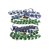
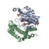
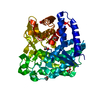
- PDB-4nez: Crystal Structure of an engineered protein with ferredoxin fold, ... -
+ Open data

- Basic information
| Entry | Database: PDB / ID: 4nez | ||||||
|---|---|---|---|---|---|---|---|
| Title | Crystal Structure of an engineered protein with ferredoxin fold, Northeast Structural Genomics Consortium (NESG) Target OR276 | ||||||
 Components Components | Engineered protein OR276 | ||||||
 Keywords Keywords | DE NOVO PROTEIN / protein engineering / ferredoxin fold / structural Genomics / PSI-Biology / Protein Structure Initiative / Northeast Structural Genomics Consortium / NESG | ||||||
| Function / homology | Ribosomal protein S10 / Alpha-Beta Plaits / 2-Layer Sandwich / Alpha Beta / tetrabutylphosphonium Function and homology information Function and homology information | ||||||
| Biological species | artificial gene (others) | ||||||
| Method |  X-RAY DIFFRACTION / SYNCHROTRON / SAD / Resolution: 2.395 Å X-RAY DIFFRACTION / SYNCHROTRON / SAD / Resolution: 2.395 Å | ||||||
 Authors Authors | Guan, R. / Lin, Y.-R. / Koga, N. / Koga, R. / Castellanos, J. / Seetharaman, J. / Maglaqui, M. / Sahdev, S. / Mao, L. / Xiao, R. ...Guan, R. / Lin, Y.-R. / Koga, N. / Koga, R. / Castellanos, J. / Seetharaman, J. / Maglaqui, M. / Sahdev, S. / Mao, L. / Xiao, R. / Everett, J.K. / Baker, D. / Montelione, G.T. / Northeast Structural Genomics Consortium (NESG) | ||||||
 Citation Citation | Journal: To be published Title: Northeast Structural Genomics Consortium Target OR276 Authors: Guan, R. / Lin, Y.-R. / Koga, N. / Koga, R. / Castellanos, J. / Seetharaman, J. / Maglaqui, M. / Sahdev, S. / Mao, L. / Xiao, R. / Everett, J.K. / Baker, D. / Montelione, G.T. / Northeast ...Authors: Guan, R. / Lin, Y.-R. / Koga, N. / Koga, R. / Castellanos, J. / Seetharaman, J. / Maglaqui, M. / Sahdev, S. / Mao, L. / Xiao, R. / Everett, J.K. / Baker, D. / Montelione, G.T. / Northeast Structural Genomics Consortium (NESG) | ||||||
| History |
|
- Structure visualization
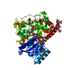
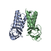
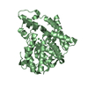
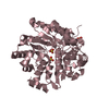
| Structure viewer | Molecule: MolmilJmol/JSmol |
|---|
- Downloads & links
-Download
| PDBx/mmCIF format | 4nez.cif.gz | 82.1 KB | Display | PDBx/mmCIF format |
|---|---|---|---|---|
| PDB format | pdb4nez.ent.gz | 62.8 KB | Display | PDB format |
| PDBx/mmJSON format | 4nez.json.gz | Tree view | PDBx/mmJSON format | |
| Others |  Other downloads Other downloads |
-Validation report
| Arichive directory | https://data.pdbj.org/pub/pdb/validation_reports/ne/4nezftp://data.pdbj.org/pub/pdb/validation_reports/ne/4nez | HTTPS FTP |
|---|
-Related structure data
| Similar structure data | |
|---|---|
| Other databases |


-Links
 PDBj
PDBj
- Assembly
| Deposited unit | 
| ||||||||||||||||||
|---|---|---|---|---|---|---|---|---|---|---|---|---|---|---|---|---|---|---|---|
| 1 | 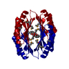
| ||||||||||||||||||
| Unit cell |
| ||||||||||||||||||
| Components on special symmetry positions |
| ||||||||||||||||||
| Details | dimer,38.91 kD,97.8% |
-Components
| #1: Protein | Mass: 20369.045 Da / Num. of mol.: 1 / Source method: obtained synthetically / Details: This is a designed protein. / Source: (synth.) artificial gene (others) |
|---|---|
| #2: Chemical | ChemComp-TRS /   Mass: 122.143 Da / Num. of mol.: 1 / Source method: obtained synthetically / Formula: C4H12NO3 / Comment: pH buffer*YM Mass: 122.143 Da / Num. of mol.: 1 / Source method: obtained synthetically / Formula: C4H12NO3 / Comment: pH buffer*YM |
| #3: Chemical | ChemComp-4NE / 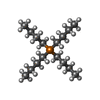  Mass: 259.431 Da / Num. of mol.: 1 / Source method: obtained synthetically / Formula: C16H36P Mass: 259.431 Da / Num. of mol.: 1 / Source method: obtained synthetically / Formula: C16H36P |
| #4: Water | ChemComp-HOH /  Mass: 18.015 Da / Num. of mol.: 20 / Source method: isolated from a natural source / Formula: H2O Mass: 18.015 Da / Num. of mol.: 20 / Source method: isolated from a natural source / Formula: H2O |
| Has protein modification | Y |
-Experimental details
-Experiment
| Experiment | Method: X-RAY DIFFRACTION / Number of used crystals: 1 |
|---|
- Sample preparation
| Crystal | Density Matthews: 2.48 Å3/Da / Density % sol: 50.47 % |
|---|---|
| Crystal grow | Temperature: 277 K / Method: vapor diffusion, hanging drop / pH: 7.4 Details: Protein solution - 100mM NaCl, 5mM DTT, 0.02% NaN3, 10mM Tris-HCl (pH 7.5), Reservoir solution - 25% PEG 3350, 0.1 M Hepes pH 7.4, 5% w/v Tetrabutylphosphonium bromide, VAPOR DIFFUSION, ...Details: Protein solution - 100mM NaCl, 5mM DTT, 0.02% NaN3, 10mM Tris-HCl (pH 7.5), Reservoir solution - 25% PEG 3350, 0.1 M Hepes pH 7.4, 5% w/v Tetrabutylphosphonium bromide, VAPOR DIFFUSION, HANGING DROP, temperature 277K |
-Data collection
| Diffraction | Mean temperature: 100 K |
|---|---|
| Diffraction source | Source: SYNCHROTRON / Site: NSLS  / Beamline: X4C / Wavelength: 0.97924 Å / Beamline: X4C / Wavelength: 0.97924 Å |
| Detector | Type: MAR CCD 165 mm / Detector: CCD / Date: May 28, 2013 |
| Radiation | Monochromator: Si 111 CHANNEL / Protocol: SINGLE WAVELENGTH / Monochromatic (M) / Laue (L): M / Scattering type: x-ray |
| Radiation wavelength | Wavelength: 0.97924 Å / Relative weight: 1 |
| Reflection | Resolution: 2.39→50 Å / Num. all: 8323 / Num. obs: 8240 / % possible obs: 99 % / Observed criterion σ(F): 2 / Observed criterion σ(I): 2 / Redundancy: 13.9 % / Biso Wilson estimate: 64.93 Å2 / Rmerge(I) obs: 0.076 |
| Reflection shell | Resolution: 2.39→2.48 Å / Redundancy: 9.9 % / Rmerge(I) obs: 0.516 / % possible all: 96.9 |
- Processing
| Software |
| |||||||||||||||||||||||||||||||||||||||||||||||||
|---|---|---|---|---|---|---|---|---|---|---|---|---|---|---|---|---|---|---|---|---|---|---|---|---|---|---|---|---|---|---|---|---|---|---|---|---|---|---|---|---|---|---|---|---|---|---|---|---|---|---|
| Refinement | Method to determine structure: SAD / Resolution: 2.395→27.566 Å / Occupancy max: 1 / Occupancy min: 0.46 / SU ML: 0.29 / σ(F): 0 / Phase error: 28.9 / Stereochemistry target values: MLHL
| |||||||||||||||||||||||||||||||||||||||||||||||||
| Solvent computation | Shrinkage radii: 0.9 Å / VDW probe radii: 1.11 Å / Solvent model: FLAT BULK SOLVENT MODEL | |||||||||||||||||||||||||||||||||||||||||||||||||
| Displacement parameters | Biso mean: 94.992 Å2 | |||||||||||||||||||||||||||||||||||||||||||||||||
| Refinement step | Cycle: LAST / Resolution: 2.395→27.566 Å
| |||||||||||||||||||||||||||||||||||||||||||||||||
| Refine LS restraints |
| |||||||||||||||||||||||||||||||||||||||||||||||||
| LS refinement shell |
| |||||||||||||||||||||||||||||||||||||||||||||||||
| Refinement TLS params. | Method: refined / Origin x: 7.7953 Å / Origin y: 40.7593 Å / Origin z: 30.5393 Å
| |||||||||||||||||||||||||||||||||||||||||||||||||
| Refinement TLS group | Selection details: chain A |
