 Movie
Movie Controller


+ Open data

- Basic information
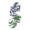
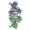
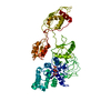
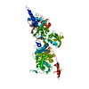
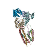
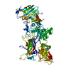
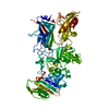
| Entry | Database: PDB / ID: 1dar | ||||||
|---|---|---|---|---|---|---|---|
| Title | ELONGATION FACTOR G IN COMPLEX WITH GDP | ||||||
 Components Components | ELONGATION FACTOR G | ||||||
 Keywords Keywords | TRANSLATIONAL GTPASE / RIBOSOMAL TRANSLOCASE | ||||||
| Function / homology |  Function and homology information Function and homology informationribosome disassembly / translation elongation factor activity / GTPase activity / GTP binding / cytoplasm Similarity search - Function | ||||||
| Biological species |   Thermus thermophilus (bacteria) Thermus thermophilus (bacteria) | ||||||
| Method |  X-RAY DIFFRACTION / SYNCHROTRON / Resolution: 2.4 Å X-RAY DIFFRACTION / SYNCHROTRON / Resolution: 2.4 Å | ||||||
 Authors Authors | Al-Karadaghi, S. / Aevarsson, A. / Garber, M. / Zheltonosova, J. / Liljas, A. | ||||||
 Citation Citation | Journal: Structure / Year: 1996 Title: The structure of elongation factor G in complex with GDP: conformational flexibility and nucleotide exchange. Authors: al-Karadaghi, S. / Aevarsson, A. / Garber, M. / Zheltonosova, J. / Liljas, A. #1: Journal: Embo J. / Year: 1994Title: Three-Dimensional Structure of the Ribosomal Translocase: Elongation Factor G from Thermus Thermophilus Authors: Aevarsson, A. / Brazhnikov, E. / Garber, M. / Zheltonosova, J. / Chirgadze, Y. / Al-Karadaghi, S. / Svensson, L.A. / Liljas, A. #2: Journal: Embo J. / Year: 1994Title: The Crystal Structure of Elongation Factor G Complexed with Gdp, at 2.7 A Resolution Authors: Czworkowski, J. / Wang, J. / Steitz, T.A. / Moore, P.B. | ||||||
| History |
|
- Structure visualization
| Structure viewer | Molecule: MolmilJmol/JSmol |
|---|
- Downloads & links
-Download
| PDBx/mmCIF format | 1dar.cif.gz | 137.4 KB | Display | PDBx/mmCIF format |
|---|---|---|---|---|
| PDB format | pdb1dar.ent.gz | 105.7 KB | Display | PDB format |
| PDBx/mmJSON format | 1dar.json.gz | Tree view | PDBx/mmJSON format | |
| Others |  Other downloads Other downloads |
-Validation report
| Arichive directory | https://data.pdbj.org/pub/pdb/validation_reports/da/1darftp://data.pdbj.org/pub/pdb/validation_reports/da/1dar | HTTPS FTP |
|---|
-Related structure data
| Similar structure data |
|---|


-Links
 PDBj
PDBj


- Assembly
| Deposited unit | 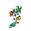
| ||||||||
|---|---|---|---|---|---|---|---|---|---|
| 1 |
| ||||||||
| Unit cell |
|
-Components
| #1: Protein | Mass: 76977.102 Da / Num. of mol.: 1 / Source method: isolated from a natural source / Source: (natural) Thermus thermophilus (bacteria) / Strain: HB8 / References: UniProt: Q5SHN5 |
|---|---|
| #2: Chemical | ChemComp-GDP /   Type: RNA linking / Mass: 443.201 Da / Num. of mol.: 1 / Source method: obtained synthetically / Formula: C10H15N5O11P2 / Comment: GDP, energy-carrying molecule*YM Type: RNA linking / Mass: 443.201 Da / Num. of mol.: 1 / Source method: obtained synthetically / Formula: C10H15N5O11P2 / Comment: GDP, energy-carrying molecule*YM |
| #3: Water | ChemComp-HOH /  Mass: 18.015 Da / Num. of mol.: 133 / Source method: isolated from a natural source / Formula: H2O Mass: 18.015 Da / Num. of mol.: 133 / Source method: isolated from a natural source / Formula: H2O |
-Experimental details
-Experiment
| Experiment | Method: X-RAY DIFFRACTION / Number of used crystals: 3 |
|---|
- Sample preparation
| Crystal | Density Matthews: 3.05 Å3/Da / Density % sol: 59.62 % | |||||||||||||||||||||||||
|---|---|---|---|---|---|---|---|---|---|---|---|---|---|---|---|---|---|---|---|---|---|---|---|---|---|---|
| Crystal grow | pH: 3 / Details: pH 3. | |||||||||||||||||||||||||
| Crystal grow | *PLUS Temperature: 4 ℃ / pH: 7.8 / Method: vapor diffusion, hanging drop / Details: Reshetnikova, L. S., (1983) FEBS Lett., 154, 149. | |||||||||||||||||||||||||
| Components of the solutions | *PLUS
|
-Data collection
| Diffraction | Mean temperature: 295 K |
|---|---|
| Diffraction source | Source: SYNCHROTRON / Site: SRS  / Beamline: PX9.6 / Wavelength: 0.87 / Beamline: PX9.6 / Wavelength: 0.87 |
| Detector | Type: MARRESEARCH / Detector: IMAGE PLATE / Date: May 1, 1994 |
| Radiation | Monochromatic (M) / Laue (L): M / Scattering type: x-ray |
| Radiation wavelength | Wavelength: 0.87 Å / Relative weight: 1 |
| Reflection | Resolution: 2.4→10 Å / Num. obs: 32595 / % possible obs: 87 % / Observed criterion σ(I): 3 / Redundancy: 4.2 % / Rmerge(I) obs: 0.07 |
| Reflection shell | Highest resolution: 2.4 Å / Rmerge(I) obs: 0.26 / Mean I/σ(I) obs: 2.9 / % possible all: 46 |
| Reflection | *PLUS Num. measured all: 284145 |
| Reflection shell | *PLUS % possible obs: 46 % |
- Processing
| Software |
| ||||||||||||||||||||||||||||||||||||||||||||||||||||||||||||
|---|---|---|---|---|---|---|---|---|---|---|---|---|---|---|---|---|---|---|---|---|---|---|---|---|---|---|---|---|---|---|---|---|---|---|---|---|---|---|---|---|---|---|---|---|---|---|---|---|---|---|---|---|---|---|---|---|---|---|---|---|---|
| Refinement | Resolution: 2.4→8 Å / σ(F): 0 Details: ATOMS WITH ZERO OCCUPANCY COULD NOT BE LOCATED IN THE ELECTRON DENSITY MAP. RESIDUES 39 - 66 (THE EFFECTOR LOOP) COULD NOT BE LOCALIZED IN THE ELECTRON DENSITY MAP. DOMAIN III (APPROXIMATELY ...Details: ATOMS WITH ZERO OCCUPANCY COULD NOT BE LOCATED IN THE ELECTRON DENSITY MAP. RESIDUES 39 - 66 (THE EFFECTOR LOOP) COULD NOT BE LOCALIZED IN THE ELECTRON DENSITY MAP. DOMAIN III (APPROXIMATELY RESIDUES 400 - 480) IS DISORDERED. SOME PARTS WERE MODELED AS POLYALANINE. THESE INCLUDE RESIDUES 433 - 442, 448 - 456, 466 - 476.
| ||||||||||||||||||||||||||||||||||||||||||||||||||||||||||||
| Refinement step | Cycle: LAST / Resolution: 2.4→8 Å
| ||||||||||||||||||||||||||||||||||||||||||||||||||||||||||||
| Refine LS restraints |
| ||||||||||||||||||||||||||||||||||||||||||||||||||||||||||||
| Software | *PLUS Name: X-PLOR / Version: 3.1 / Classification: refinement | ||||||||||||||||||||||||||||||||||||||||||||||||||||||||||||
| Refinement | *PLUS Rfactor obs: 0.22 / Rfactor Rfree: 0.29 / Rfactor Rwork: 0.22 | ||||||||||||||||||||||||||||||||||||||||||||||||||||||||||||
| Solvent computation | *PLUS | ||||||||||||||||||||||||||||||||||||||||||||||||||||||||||||
| Displacement parameters | *PLUS | ||||||||||||||||||||||||||||||||||||||||||||||||||||||||||||
| Refine LS restraints | *PLUS
|
