Mass: 1723.917 Da / Num. of mol.: 1 / Source method: obtained synthetically Details: HISTIDINE TAG IS ENGINEERED INTO THE VECTOR THAT IS EXPRESSED WITH THE PROTEIN
#2: Protein
TRANSLATIONINITIATIONFACTOR1A
Mass: 16357.254 Da / Num. of mol.: 1 Source method: isolated from a genetically manipulated source Source: (gene. exp.) Homo sapiens (human) / Plasmid: PQE31 / Production host: Escherichia coli (E. coli) / References: UniProt: P47813
-
Experimental details
-
Experiment
Experiment
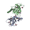
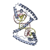
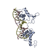
Method: SOLUTION NMR
NMR experiment
Conditions-ID
Experiment-ID
Solution-ID
Type
1
1
1
3D 13C-SEPARATED NOESY
2
2
2
3D 15N-SEPARATED NOESY
3
3
3
2D NOESY
NMR details
Text: THE PROTEIN WAS ASSIGNED FROM THE TRIPLE RESONANCE EXPERIMENTS HNCA, HN(CO)CA, HNCO, HN(CA)CO, CBCA(CO)NH, HBHA(CBCACO)NH, H(CCCO)NH, (H)C(CCO)NH, AND HCCH- TOCSY. VAL AND LEU METHYL GROUPS ...Text: THE PROTEIN WAS ASSIGNED FROM THE TRIPLE RESONANCE EXPERIMENTS HNCA, HN(CO)CA, HNCO, HN(CA)CO, CBCA(CO)NH, HBHA(CBCACO)NH, H(CCCO)NH, (H)C(CCO)NH, AND HCCH- TOCSY. VAL AND LEU METHYL GROUPS WERE STEREOSPECIFICALLY ASSIGNED FROM ANALYSIS OF MULTIPLET COUPLINGS IN AN HSQC OF 10% 13C-LABELED PROTEIN. BACKBONE DIHEDRAL ANGLES WERE ESTIMATED USING THE PROGRAM TALOS AND BACKBONE HETERONUCLEAR CHEMICAL SHIFTS.
C. BARTELS, T. XIA, M. BILLETER, P. GUNTERT, ANDK. WUTHRICH
dataanalysis
X-PLOR
3.851
A.T. BRUNGER
structuresolution
TALOS
1
G. CORNILESCU, F. DELAGLIO, ANDA. BAX
dataanalysis
X-PLOR
3.851
A.T. BRUNGER
refinement
Refinement
Method: STRUCTURES WERE CALCULATED USING SIMULATED ANNEALING, MOLECULAR DYNAMICS FROM AN EXTENDED CONFORMATION WITH RANDOM PHI, PSI ANGLES. Software ordinal: 1 Details: CALCULATIONS WERE RESTRAINED WITH THE FOLLOWING NMR DATA. NOE DISTANCE RESTRAINTS INTRA-RESIDUE 607 INTER-RESIDUE SEQUENTIAL (IJ=1) 266 INTER-RESIDUE MEDIUM (IJ 4) 342 HYDROGENS BONDS: ...Details: CALCULATIONS WERE RESTRAINED WITH THE FOLLOWING NMR DATA. NOE DISTANCE RESTRAINTS INTRA-RESIDUE 607 INTER-RESIDUE SEQUENTIAL (IJ=1) 266 INTER-RESIDUE MEDIUM (IJ <= 4) 105 INTER-RESIDUE LONG (IJ > 4) 342 HYDROGENS BONDS: PROTEIN BACKBONE 40 DIHEDRAL ANGLES: PHI 65 PSI 65 NO NOE VIOLATIONS >0.3 A IN FINAL STRUCTURES. RMS DEVIATIONS OF THE ENSEMBLE SUPERPOSITION TO THE AVERAGE STRUCTURE ARE: BACKBONE 39-131 0.57 A SECONDARY STRUCTURE 0.23 A HEAVY ATOMS 39-131 1.17 A SECONDARY STRUCTURE 0.72 A BOND LENGTHS 0.0016 A BOND ANGLES 0.345 DEGREE IMPROPER ANGLES 0.259 DEGREE. NOTE THAT RESIDUES 1-38 AND 132-143 HAD NO LONG RANGE NOES AND ARE DISORDERED IN THE CALCULATED STRUCTURES.
NMR representative
Selection criteria: lowest energy
NMR ensemble
Conformer selection criteria: structures with the least restraint violations Conformers calculated total number: 50 / Conformers submitted total number: 20
+
About Yorodumi
-
News
-
Feb 9, 2022. New format data for meta-information of EMDB entries
New format data for meta-information of EMDB entries
Version 3 of the EMDB header file is now the official format.
The previous official version 1.9 will be removed from the archive.
In the structure databanks used in Yorodumi, some data are registered as the other names, "COVID-19 virus" and "2019-nCoV". Here are the details of the virus and the list of structure data.
Jan 31, 2019. EMDB accession codes are about to change! (news from PDBe EMDB page)
EMDB accession codes are about to change! (news from PDBe EMDB page)
The allocation of 4 digits for EMDB accession codes will soon come to an end. Whilst these codes will remain in use, new EMDB accession codes will include an additional digit and will expand incrementally as the available range of codes is exhausted. The current 4-digit format prefixed with “EMD-” (i.e. EMD-XXXX) will advance to a 5-digit format (i.e. EMD-XXXXX), and so on. It is currently estimated that the 4-digit codes will be depleted around Spring 2019, at which point the 5-digit format will come into force.
The EM Navigator/Yorodumi systems omit the EMD- prefix.
Related info.:Q: What is EMD? / ID/Accession-code notation in Yorodumi/EM Navigator
Yorodumi is a browser for structure data from EMDB, PDB, SASBDB, etc.
This page is also the successor to EM Navigator detail page, and also detail information page/front-end page for Omokage search.
The word "yorodu" (or yorozu) is an old Japanese word meaning "ten thousand". "mi" (miru) is to see.
Related info.:EMDB / PDB / SASBDB / Comparison of 3 databanks / Yorodumi Search / Aug 31, 2016. New EM Navigator & Yorodumi / Yorodumi Papers / Jmol/JSmol / Function and homology information / Changes in new EM Navigator and Yorodumi
 Movie
Movie Controller


 Open data
 Basic information
 Components
 Keywords
 Function and homology information
 Homo sapiens (human)
 Authors
 Citation
 Structure visualization
 Downloads & links
 Other downloads
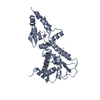


 PDBj
PDBj


 Assembly

 Sample preparation
 Processing
 X-PLOR