 Movie
Movie Controller

[English] 日本語
 Yorodumi
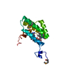
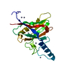
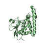
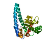
- PDB-1jod: Crystal Structure of Murine Olfactory Marker Protein in Spacegrou... -
+ Open data

- Basic information
| Entry | Database: PDB / ID: 1jod | ||||||
|---|---|---|---|---|---|---|---|
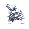
| Title | Crystal Structure of Murine Olfactory Marker Protein in Spacegroup P43212 | ||||||
 Components Components | Olfactory Marker Protein | ||||||
 Keywords Keywords | SIGNALING PROTEIN / Beta Clamshell | ||||||
| Function / homology |  Function and homology information Function and homology informationdendritic knob / peptide binding / neurogenesis / acrosomal vesicle / sensory perception of smell / sperm midpiece / axon / neuronal cell body / dendrite / signal transduction ...dendritic knob / peptide binding / neurogenesis / acrosomal vesicle / sensory perception of smell / sperm midpiece / axon / neuronal cell body / dendrite / signal transduction / nucleus / cytoplasm / cytosol Similarity search - Function | ||||||
| Biological species |  | ||||||
| Method |  X-RAY DIFFRACTION / SYNCHROTRON / MOLECULAR REPLACEMENT / Resolution: 3.2 Å X-RAY DIFFRACTION / SYNCHROTRON / MOLECULAR REPLACEMENT / Resolution: 3.2 Å | ||||||
 Authors Authors | Smith, P. / Hunt, J.F. | ||||||
 Citation Citation | Journal: J.Mol.Biol. / Year: 2002 Title: The crystal structure of the olfactory marker protein at 2.3 A resolution. Authors: Smith, P.C. / Firestein, S. / Hunt, J.F. | ||||||
| History |
|
- Structure visualization
| Structure viewer | Molecule: MolmilJmol/JSmol |
|---|
- Downloads & links
-Download
| PDBx/mmCIF format | 1jod.cif.gz | 82.6 KB | Display | PDBx/mmCIF format |
|---|---|---|---|---|
| PDB format | pdb1jod.ent.gz | 61.1 KB | Display | PDB format |
| PDBx/mmJSON format | 1jod.json.gz | Tree view | PDBx/mmJSON format | |
| Others |  Other downloads Other downloads |
-Validation report
| Arichive directory | https://data.pdbj.org/pub/pdb/validation_reports/jo/1jodftp://data.pdbj.org/pub/pdb/validation_reports/jo/1jod | HTTPS FTP |
|---|
-Related structure data
| Related structure data |  1f35C  1jobC  1f15S C: citing same article ( S: Starting model for refinement |
|---|---|
| Similar structure data |


-Links
 PDBj
PDBj


- Assembly
| Deposited unit | 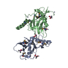
| ||||||||
|---|---|---|---|---|---|---|---|---|---|
| 1 | 
| ||||||||
| 2 | 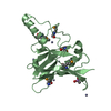
| ||||||||
| Unit cell |
|
-Components
| #1: Protein | Mass: 18991.777 Da / Num. of mol.: 2 Source method: isolated from a genetically manipulated source Source: (gene. exp.)  #2: Chemical | ChemComp-ZN /   Mass: 65.409 Da / Num. of mol.: 22 / Source method: obtained synthetically / Formula: Zn Mass: 65.409 Da / Num. of mol.: 22 / Source method: obtained synthetically / Formula: Zn#3: Chemical | ChemComp-CAC /   Mass: 136.989 Da / Num. of mol.: 4 / Source method: obtained synthetically / Formula: C2H6AsO2 Mass: 136.989 Da / Num. of mol.: 4 / Source method: obtained synthetically / Formula: C2H6AsO2#4: Water | ChemComp-HOH / |  Mass: 18.015 Da / Num. of mol.: 31 / Source method: isolated from a natural source / Formula: H2O Mass: 18.015 Da / Num. of mol.: 31 / Source method: isolated from a natural source / Formula: H2OHas protein modification | Y | |
|---|
-Experimental details
-Experiment
| Experiment | Method: X-RAY DIFFRACTION / Number of used crystals: 1 |
|---|
- Sample preparation
| Crystal | Density Matthews: 4.15 Å3/Da / Density % sol: 70.39 % |
|---|---|
| Crystal grow | Temperature: 295 K / Method: vapor diffusion, hanging drop / pH: 6.5 Details: PEG 8000, Zinc Acetate, Cacodylate, pH 6.5, VAPOR DIFFUSION, HANGING DROP, temperature 295K |
| Crystal grow | *PLUS Method: unknown / Details: unpublished structure |
-Data collection
| Diffraction | Mean temperature: 130 K |
|---|---|
| Diffraction source | Source: SYNCHROTRON / Site: NSLS  / Beamline: X12C / Wavelength: 0.9785 Å / Beamline: X12C / Wavelength: 0.9785 Å |
| Detector | Type: CUSTOM-MADE / Detector: CCD / Date: Apr 15, 2000 |
| Radiation | Monochromator: Si 111 CHANNEL / Protocol: SINGLE WAVELENGTH / Monochromatic (M) / Laue (L): M / Scattering type: x-ray |
| Radiation wavelength | Wavelength: 0.9785 Å / Relative weight: 1 |
| Reflection | Resolution: 3.2→40 Å / Num. all: 11102 / Num. obs: 8962 / % possible obs: 80.4 % / Observed criterion σ(F): 1 / Observed criterion σ(I): -3 / Biso Wilson estimate: 59.1 Å2 |
| Reflection shell | Resolution: 3.2→3.31 Å / % possible all: 61.9 |
- Processing
| Software |
| |||||||||||||||||||||||||
|---|---|---|---|---|---|---|---|---|---|---|---|---|---|---|---|---|---|---|---|---|---|---|---|---|---|---|
| Refinement | Method to determine structure: MOLECULAR REPLACEMENT Starting model: PDB ID 1F15 Resolution: 3.2→40 Å / Rfactor Rfree error: 0.011 / Data cutoff high absF: 1000000 / Data cutoff low absF: 0.01 / Isotropic thermal model: RESTRAINED / Cross valid method: THROUGHOUT / σ(F): 2 / Stereochemistry target values: Engh & Huber
| |||||||||||||||||||||||||
| Displacement parameters | Biso mean: 28.6 Å2
| |||||||||||||||||||||||||
| Refine analyze |
| |||||||||||||||||||||||||
| Refinement step | Cycle: LAST / Resolution: 3.2→40 Å
| |||||||||||||||||||||||||
| Refine LS restraints |
| |||||||||||||||||||||||||
| LS refinement shell | Resolution: 3.2→3.31 Å / Rfactor Rfree error: 0.045 / Total num. of bins used: 10
|
