 Movie
Movie Controller


+ Open data

- Basic information
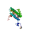
| Entry | Database: PDB / ID: 1f35 | ||||||
|---|---|---|---|---|---|---|---|
| Title | CRYSTAL STRUCTURE OF MURINE OLFACTORY MARKER PROTEIN | ||||||
 Components Components | OLFACTORY MARKER PROTEIN | ||||||
 Keywords Keywords | SIGNALING PROTEIN / BETA / Structural Genomics / PSI / Protein Structure Initiative / Northeast Structural Genomics Consortium / NESG | ||||||
| Function / homology |  Function and homology information Function and homology informationdendritic knob / peptide binding / neurogenesis / acrosomal vesicle / sensory perception of smell / sperm midpiece / axon / neuronal cell body / dendrite / signal transduction ...dendritic knob / peptide binding / neurogenesis / acrosomal vesicle / sensory perception of smell / sperm midpiece / axon / neuronal cell body / dendrite / signal transduction / nucleus / cytoplasm / cytosol Similarity search - Function | ||||||
| Biological species |  | ||||||
| Method |  X-RAY DIFFRACTION / SYNCHROTRON / MAD / Resolution: 2.3 Å X-RAY DIFFRACTION / SYNCHROTRON / MAD / Resolution: 2.3 Å | ||||||
 Authors Authors | Smith, P.C. / Hunt, J.F. / Northeast Structural Genomics Consortium (NESG) | ||||||
 Citation Citation | Journal: J.Mol.Biol. / Year: 2002 Title: The crystal structure of the olfactory marker protein at 2.3 A resolution. Authors: Smith, P.C. / Firestein, S. / Hunt, J.F. | ||||||
| History |
|
- Structure visualization
| Structure viewer | Molecule: MolmilJmol/JSmol |
|---|
- Downloads & links
-Download
| PDBx/mmCIF format | 1f35.cif.gz | 85.1 KB | Display | PDBx/mmCIF format |
|---|---|---|---|---|
| PDB format | pdb1f35.ent.gz | 63.5 KB | Display | PDB format |
| PDBx/mmJSON format | 1f35.json.gz | Tree view | PDBx/mmJSON format | |
| Others |  Other downloads Other downloads |
-Validation report
| Arichive directory | https://data.pdbj.org/pub/pdb/validation_reports/f3/1f35ftp://data.pdbj.org/pub/pdb/validation_reports/f3/1f35 | HTTPS FTP |
|---|
-Related structure data


-Links
 PDBj
PDBj


- Assembly
| Deposited unit | 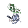
| ||||||||
|---|---|---|---|---|---|---|---|---|---|
| 1 | 
| ||||||||
| 2 | 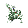
| ||||||||
| 3 | 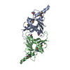
| ||||||||
| Unit cell |
| ||||||||
| Details | The biological assembly is a monomer. |
-Components
| #1: Protein | Mass: 18991.777 Da / Num. of mol.: 2 Mutation: M13(MSE), M25(MSE), M95(MSE), M115(MSE), M139(MSE) Source method: isolated from a genetically manipulated source Source: (gene. exp.)  #2: Chemical | ChemComp-ZN /   Mass: 65.409 Da / Num. of mol.: 10 / Source method: obtained synthetically / Formula: Zn Mass: 65.409 Da / Num. of mol.: 10 / Source method: obtained synthetically / Formula: Zn#3: Chemical | ChemComp-CAC / |   Mass: 136.989 Da / Num. of mol.: 1 / Source method: obtained synthetically / Formula: C2H6AsO2 Mass: 136.989 Da / Num. of mol.: 1 / Source method: obtained synthetically / Formula: C2H6AsO2#4: Water | ChemComp-HOH / |  Mass: 18.015 Da / Num. of mol.: 176 / Source method: isolated from a natural source / Formula: H2O Mass: 18.015 Da / Num. of mol.: 176 / Source method: isolated from a natural source / Formula: H2OHas protein modification | Y | |
|---|
-Experimental details
-Experiment
| Experiment | Method: X-RAY DIFFRACTION / Number of used crystals: 1 |
|---|
- Sample preparation
| Crystal | Density Matthews: 4.75 Å3/Da / Density % sol: 74.09 % Description: 4 wavelengths used were: peak: 0.9786, edge: 0.9788, high energy remote: 0.9390, low energy remote: 1.078. | |||||||||||||||||||||||||
|---|---|---|---|---|---|---|---|---|---|---|---|---|---|---|---|---|---|---|---|---|---|---|---|---|---|---|
| Crystal grow | Temperature: 294 K / Method: vapor diffusion, hanging drop / pH: 6.5 Details: PEG 8000, Zinc Acetate, Cacodylate, pH 6.5, VAPOR DIFFUSION, HANGING DROP, temperature 21K | |||||||||||||||||||||||||
| Crystal grow | *PLUS | |||||||||||||||||||||||||
| Components of the solutions | *PLUS
|
-Data collection
| Diffraction | Mean temperature: 130 K | |||||||||||||||
|---|---|---|---|---|---|---|---|---|---|---|---|---|---|---|---|---|
| Diffraction source | Source: SYNCHROTRON / Site: NSLS  / Beamline: X12C / Wavelength: 0.9786, 0.9788, 0.9390, 1.078 / Beamline: X12C / Wavelength: 0.9786, 0.9788, 0.9390, 1.078 | |||||||||||||||
| Detector | Type: BRANDEIS / Detector: CCD / Date: Apr 20, 2000 | |||||||||||||||
| Radiation | Protocol: MAD / Monochromatic (M) / Laue (L): M / Scattering type: x-ray | |||||||||||||||
| Radiation wavelength |
| |||||||||||||||
| Reflection | Resolution: 2.2→40 Å / Num. all: 48740 / Num. obs: 35024 / % possible obs: 74 % / Observed criterion σ(F): 1 / Observed criterion σ(I): 3 / Redundancy: 16.6 % / Biso Wilson estimate: 41.6 Å2 / Rmerge(I) obs: 0.058 / Net I/σ(I): 2.69 | |||||||||||||||
| Reflection shell | Resolution: 2.3→2.4 Å / Redundancy: 16.4 % / Rmerge(I) obs: 0.25 / Num. unique all: 2766 / % possible all: 71.8 |
- Processing
| Software |
| |||||||||||||||||||||||||
|---|---|---|---|---|---|---|---|---|---|---|---|---|---|---|---|---|---|---|---|---|---|---|---|---|---|---|
| Refinement | Method to determine structure: MAD / Resolution: 2.3→100 Å / Rfactor Rfree error: 0.007 / Data cutoff high absF: 1000000 / Data cutoff low absF: 0.01 / Isotropic thermal model: RESTRAINED / Cross valid method: THROUGHOUT / σ(F): 1 / σ(I): 1 / Stereochemistry target values: X-PLOR 3.851
| |||||||||||||||||||||||||
| Displacement parameters | Biso mean: 49.3 Å2
| |||||||||||||||||||||||||
| Refine analyze |
| |||||||||||||||||||||||||
| Refinement step | Cycle: LAST / Resolution: 2.3→100 Å
| |||||||||||||||||||||||||
| Refine LS restraints |
| |||||||||||||||||||||||||
| LS refinement shell | Resolution: 2.3→2.4 Å / Rfactor Rfree error: 0.034 / Total num. of bins used: 8
| |||||||||||||||||||||||||
| Xplor file |
| |||||||||||||||||||||||||
| Software | *PLUS Name: X-PLOR / Version: 3.851 / Classification: refinement | |||||||||||||||||||||||||
| Refinement | *PLUS Lowest resolution: 100 Å / σ(F): 1 / % reflection Rfree: 5 % / Rfactor obs: 0.21 / Rfactor Rwork: 0.21 | |||||||||||||||||||||||||
| Solvent computation | *PLUS | |||||||||||||||||||||||||
| Displacement parameters | *PLUS Biso mean: 49.3 Å2 | |||||||||||||||||||||||||
| Refine LS restraints | *PLUS
| |||||||||||||||||||||||||
| LS refinement shell | *PLUS Rfactor Rfree: 0.373 / % reflection Rfree: 4.3 % / Rfactor Rwork: 0.349 |
