 Movie
Movie Controller

[English] 日本語
 Yorodumi
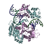
- PDB-1d7k: CRYSTAL STRUCTURE OF HUMAN ORNITHINE DECARBOXYLASE AT 2.1 ANGSTRO... -
+ Open data

- Basic information
| Entry | Database: PDB / ID: 1d7k | ||||||
|---|---|---|---|---|---|---|---|
| Title | CRYSTAL STRUCTURE OF HUMAN ORNITHINE DECARBOXYLASE AT 2.1 ANGSTROMS RESOLUTION | ||||||
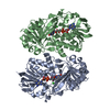
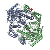
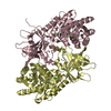
 Components Components | HUMAN ORNITHINE DECARBOXYLASE | ||||||
 Keywords Keywords | LYASE / ALPHA-BETA BARREL / PYRIDOXAL 5'-PHOSPHATE / SHEET-DOMAIN / DECARBOXYLATION / ORNITHINE | ||||||
| Function / homology |  Function and homology information Function and homology informationornithine decarboxylase / : / ornithine decarboxylase activity / Metabolism of polyamines / polyamine metabolic process / Regulation of ornithine decarboxylase (ODC) / regulation of protein catabolic process / kidney development / response to virus / cell population proliferation ...ornithine decarboxylase / : / ornithine decarboxylase activity / Metabolism of polyamines / polyamine metabolic process / Regulation of ornithine decarboxylase (ODC) / regulation of protein catabolic process / kidney development / response to virus / cell population proliferation / positive regulation of cell population proliferation / protein homodimerization activity / cytosol / cytoplasm Similarity search - Function | ||||||
| Biological species |  Homo sapiens (human) Homo sapiens (human) | ||||||
| Method |  X-RAY DIFFRACTION / SYNCHROTRON / Resolution: 2.1 Å X-RAY DIFFRACTION / SYNCHROTRON / Resolution: 2.1 Å | ||||||
 Authors Authors | Almrud, J.J. / Oliveira, M.A. / Kern, A.D. / Grishin, N.V. / Phillips, M.A. / Hackert, M.L. | ||||||
 Citation Citation | Journal: J.Mol.Biol. / Year: 2000 Title: Crystal structure of human ornithine decarboxylase at 2.1 A resolution: structural insights to antizyme binding. Authors: Almrud, J.J. / Oliveira, M.A. / Kern, A.D. / Grishin, N.V. / Phillips, M.A. / Hackert, M.L. | ||||||
| History |
|
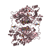
- Structure visualization
| Structure viewer | Molecule: MolmilJmol/JSmol |
|---|
- Downloads & links
-Download
| PDBx/mmCIF format | 1d7k.cif.gz | 180.4 KB | Display | PDBx/mmCIF format |
|---|---|---|---|---|
| PDB format | pdb1d7k.ent.gz | 141.3 KB | Display | PDB format |
| PDBx/mmJSON format | 1d7k.json.gz | Tree view | PDBx/mmJSON format | |
| Others |  Other downloads Other downloads |
-Validation report
| Arichive directory | https://data.pdbj.org/pub/pdb/validation_reports/d7/1d7kftp://data.pdbj.org/pub/pdb/validation_reports/d7/1d7k | HTTPS FTP |
|---|
-Related structure data
| Similar structure data |
|---|


-Links
 PDBj
PDBj
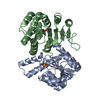
- Assembly
| Deposited unit | 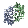
| ||||||||
|---|---|---|---|---|---|---|---|---|---|
| 1 |
| ||||||||
| Unit cell |
|
-Components
| #1: Protein | Mass: 47161.422 Da / Num. of mol.: 2 Source method: isolated from a genetically manipulated source Source: (gene. exp.) Homo sapiens (human) / Organ: LIVER / Production host:  #2: Water | ChemComp-HOH / |  Mass: 18.015 Da / Num. of mol.: 401 / Source method: isolated from a natural source / Formula: H2O Mass: 18.015 Da / Num. of mol.: 401 / Source method: isolated from a natural source / Formula: H2OHas protein modification | Y | |
|---|
-Experimental details
-Experiment
| Experiment | Method: X-RAY DIFFRACTION / Number of used crystals: 3 |
|---|
- Sample preparation
| Crystal | Density Matthews: 2.45 Å3/Da / Density % sol: 49.86 % | ||||||||||||||||||||||||||||||
|---|---|---|---|---|---|---|---|---|---|---|---|---|---|---|---|---|---|---|---|---|---|---|---|---|---|---|---|---|---|---|---|
| Crystal grow | Temperature: 289 K / Method: vapor diffusion / pH: 7.5 Details: 20% PEG 3350, 0.2M NaCl, 5mM DTT, 0.1M Tris-HCl, pH 7.5, VAPOR DIFFUSION, temperature 16K | ||||||||||||||||||||||||||||||
| Crystal grow | *PLUS Method: unknown | ||||||||||||||||||||||||||||||
| Components of the solutions | *PLUS
|
-Data collection
| Diffraction | Mean temperature: 100 K |
|---|---|
| Diffraction source | Source: SYNCHROTRON / Site: CHESS  / Beamline: A1 / Wavelength: 0.908 / Beamline: A1 / Wavelength: 0.908 |
| Detector | Type: FUJI / Detector: IMAGE PLATE / Date: Jan 15, 1996 |
| Radiation | Protocol: SINGLE WAVELENGTH / Monochromatic (M) / Laue (L): M / Scattering type: x-ray |
| Radiation wavelength | Wavelength: 0.908 Å / Relative weight: 1 |
| Reflection | Resolution: 2.1→30 Å / Num. all: 55799 / Num. obs: 53656 / % possible obs: 96.4 % / Observed criterion σ(I): 2 / Redundancy: 4 % / Biso Wilson estimate: 29 Å2 / Rmerge(I) obs: 0.068 / Net I/σ(I): 10.1 |
| Reflection shell | Resolution: 2.09→2.16 Å / Redundancy: 3.4 % / Rmerge(I) obs: 0.363 / Num. unique all: 4929 / % possible all: 97.5 |
| Reflection | *PLUS Num. measured all: 223151 |
| Reflection shell | *PLUS % possible obs: 97.5 % |
- Processing
| Software |
| |||||||||||||||||||||||||
|---|---|---|---|---|---|---|---|---|---|---|---|---|---|---|---|---|---|---|---|---|---|---|---|---|---|---|
| Refinement | Resolution: 2.1→30.5 Å / Cross valid method: throyghout / σ(F): 0 / σ(I): 0 / Stereochemistry target values: Engh & Huber Details: This structure was refined using data from 30-2.1 angstrom resolution using the CCP4 suite program REFMAC. The REFMAC refinement was carried out using the maximum likelihood function and ...Details: This structure was refined using data from 30-2.1 angstrom resolution using the CCP4 suite program REFMAC. The REFMAC refinement was carried out using the maximum likelihood function and minimization by the conjugate direction method.
| |||||||||||||||||||||||||
| Refinement step | Cycle: LAST / Resolution: 2.1→30.5 Å
| |||||||||||||||||||||||||
| Refine LS restraints |
| |||||||||||||||||||||||||
| Software | *PLUS Name: REFMAC / Classification: refinement | |||||||||||||||||||||||||
| Refine LS restraints | *PLUS
| |||||||||||||||||||||||||
| LS refinement shell | *PLUS Highest resolution: 2.1 Å / Lowest resolution: 2.2 Å / Rfactor Rfree: 0.306 / Rfactor Rwork: 0.235 / Num. reflection obs: 6009 |
