 Movie
Movie Controller

[English] 日本語
 Yorodumi
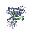
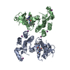
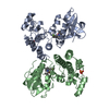
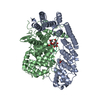
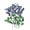
- PDB-1d3u: TATA-BINDING PROTEIN/TRANSCRIPTION FACTOR (II)B/BRE+TATA-BOX COMP... -
+ Open data

- Basic information
| Entry | Database: PDB / ID: 1d3u | ||||||
|---|---|---|---|---|---|---|---|
| Title | TATA-BINDING PROTEIN/TRANSCRIPTION FACTOR (II)B/BRE+TATA-BOX COMPLEX FROM PYROCOCCUS WOESEI | ||||||
 Components Components |
| ||||||
 Keywords Keywords | GENE REGULATION/DNA / PROTEIN-DNA COMPLEX / TRANSCRIPTION / HYPERTHERMOPHILE / GENE REGULATION-DNA COMPLEX | ||||||
| Function / homology |  Function and homology information Function and homology informationtranscription preinitiation complex assembly / transcription preinitiation complex / TBP-class protein binding / DNA-templated transcription initiation / DNA-binding transcription factor activity / DNA binding / zinc ion binding Similarity search - Function | ||||||
| Biological species |   Pyrococcus woesei (archaea) Pyrococcus woesei (archaea) | ||||||
| Method |  X-RAY DIFFRACTION / SYNCHROTRON / Resolution: 2.4 Å X-RAY DIFFRACTION / SYNCHROTRON / Resolution: 2.4 Å | ||||||
 Authors Authors | Littlefield, O. / Korkhin, Y. / Sigler, P.B. | ||||||
 Citation Citation | Journal: Proc.Natl.Acad.Sci.USA / Year: 1999 Title: The structural basis for the oriented assembly of a TBP/TFB/promoter complex. Authors: Littlefield, O. / Korkhin, Y. / Sigler, P.B. #1: Journal: Proc.Natl.Acad.Sci.USA / Year: 1999Title: The orientation of the transcription preinitiation complex in Archaea Authors: Bell, S.D. / Kosa, P.L. / Sigler, P.B. / Jackson, S.P. #2: Journal: Proc.Natl.Acad.Sci.USA / Year: 1997Title: The 2.1-angstrom crystal structure of an archaeal preinitiation complex: TATA- box-binding protein/transcription factor (II)B core/TATA-box Authors: Kosa, P.F. / Ghosh, G. / DeDecker, B.S. / Sigler, P.B. #3: Journal: Mol.Cell / Year: 1998Title: Sequence-specific DNA binding by the S. shibatae TFIIB homolog, TFB, and its effect on promoter strength Authors: Qureshi, S.A. / Jackson, S.P. #4: Journal: Genes Dev. / Year: 1998Title: New core promoter element in RNA polymerase II-dependent transcription: sequence-specific DNA binding by transcription factor IIB Authors: Lagrange, T. / Kapanidis, A.N. / Tang, H. / Reinberg, D. / Ebright, R.H. | ||||||
| History |
|
- Structure visualization
| Structure viewer | Molecule: MolmilJmol/JSmol |
|---|
- Downloads & links
-Download
| PDBx/mmCIF format | 1d3u.cif.gz | 120.3 KB | Display | PDBx/mmCIF format |
|---|---|---|---|---|
| PDB format | pdb1d3u.ent.gz | 89.3 KB | Display | PDB format |
| PDBx/mmJSON format | 1d3u.json.gz | Tree view | PDBx/mmJSON format | |
| Others |  Other downloads Other downloads |
-Validation report
| Arichive directory | https://data.pdbj.org/pub/pdb/validation_reports/d3/1d3uftp://data.pdbj.org/pub/pdb/validation_reports/d3/1d3u | HTTPS FTP |
|---|
-Related structure data
| Similar structure data |
|---|


-Links
 PDBj
PDBj


- Assembly
| Deposited unit | 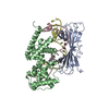
| ||||||||||
|---|---|---|---|---|---|---|---|---|---|---|---|
| 1 |
| ||||||||||
| Unit cell |
| ||||||||||
| Components on special symmetry positions |
|
-Components
| #1: DNA chain | Mass: 7414.853 Da / Num. of mol.: 1 / Source method: obtained synthetically |
|---|---|
| #2: DNA chain | Mass: 7003.568 Da / Num. of mol.: 1 / Source method: obtained synthetically |
| #3: Protein | Mass: 20166.549 Da / Num. of mol.: 1 / Fragment: RESIDUES 1-180 Source method: isolated from a genetically manipulated source Source: (gene. exp.) Pyrococcus woesei (archaea) / Plasmid: PET11A / Production host:  |
| #4: Protein | Mass: 22721.576 Da / Num. of mol.: 1 / Fragment: C-TERMINAL CORE, RESIDUES 62-261 Source method: isolated from a genetically manipulated source Source: (gene. exp.) Pyrococcus woesei (archaea) / Plasmid: PET11A / Production host: |
| #5: Water | ChemComp-HOH /  Mass: 18.015 Da / Num. of mol.: 180 / Source method: isolated from a natural source / Formula: H2O Mass: 18.015 Da / Num. of mol.: 180 / Source method: isolated from a natural source / Formula: H2O |
| Has protein modification | Y |
-Experimental details
-Experiment
| Experiment | Method: X-RAY DIFFRACTION / Number of used crystals: 1 |
|---|
- Sample preparation
| Crystal | Density Matthews: 3.84 Å3/Da / Density % sol: 68 % | ||||||||||||||||||||||||||||||||||||||||||||||||||
|---|---|---|---|---|---|---|---|---|---|---|---|---|---|---|---|---|---|---|---|---|---|---|---|---|---|---|---|---|---|---|---|---|---|---|---|---|---|---|---|---|---|---|---|---|---|---|---|---|---|---|---|
| Crystal grow | Temperature: 292 K / Method: vapor diffusion, hanging drop / pH: 8.5 Details: PEG 400, STRONTIUM CHLORIDE, SODIUM CITRATE, TRIS, pH 8.5, VAPOR DIFFUSION, HANGING DROP, temperature 292K | ||||||||||||||||||||||||||||||||||||||||||||||||||
| Components of the solutions |
| ||||||||||||||||||||||||||||||||||||||||||||||||||
| Crystal grow | *PLUS Temperature: 19 ℃ / pH: 8 | ||||||||||||||||||||||||||||||||||||||||||||||||||
| Components of the solutions | *PLUS
|
-Data collection
| Diffraction | Mean temperature: 100 K |
|---|---|
| Diffraction source | Source: SYNCHROTRON / Site: APS  / Beamline: 19-ID / Wavelength: 1.03 / Beamline: 19-ID / Wavelength: 1.03 |
| Detector | Type: CUSTOM-MADE / Detector: CCD / Date: Dec 8, 1998 |
| Radiation | Protocol: SINGLE WAVELENGTH / Monochromatic (M) / Laue (L): M / Scattering type: x-ray |
| Radiation wavelength | Wavelength: 1.03 Å / Relative weight: 1 |
| Reflection | Resolution: 2.4→28.3 Å / Num. obs: 33982 / % possible obs: 99.2 % / Observed criterion σ(F): 0 / Observed criterion σ(I): 0 / Redundancy: 5.2 % / Biso Wilson estimate: 48.2 Å2 / Rmerge(I) obs: 0.068 / Net I/σ(I): 7.3 |
| Reflection shell | Resolution: 2.4→2.53 Å / Redundancy: 4.2 % / Rmerge(I) obs: 0.339 / % possible all: 99.2 |
- Processing
| Software |
| ||||||||||||||||||||||||||||||||||||||||||||||||||||||||||||
|---|---|---|---|---|---|---|---|---|---|---|---|---|---|---|---|---|---|---|---|---|---|---|---|---|---|---|---|---|---|---|---|---|---|---|---|---|---|---|---|---|---|---|---|---|---|---|---|---|---|---|---|---|---|---|---|---|---|---|---|---|---|
| Refinement | Resolution: 2.4→20 Å / Cross valid method: THROUGHOUT / σ(F): 0 / σ(I): 0 / Stereochemistry target values: ENGH & HUBER
| ||||||||||||||||||||||||||||||||||||||||||||||||||||||||||||
| Refinement step | Cycle: LAST / Resolution: 2.4→20 Å
| ||||||||||||||||||||||||||||||||||||||||||||||||||||||||||||
| Refine LS restraints |
| ||||||||||||||||||||||||||||||||||||||||||||||||||||||||||||
| Software | *PLUS Name: CNS / Classification: refinement | ||||||||||||||||||||||||||||||||||||||||||||||||||||||||||||
| Refinement | *PLUS Highest resolution: 2.4 Å / Lowest resolution: 20 Å / σ(F): 0 / % reflection Rfree: 10 % | ||||||||||||||||||||||||||||||||||||||||||||||||||||||||||||
| Solvent computation | *PLUS | ||||||||||||||||||||||||||||||||||||||||||||||||||||||||||||
| Displacement parameters | *PLUS | ||||||||||||||||||||||||||||||||||||||||||||||||||||||||||||
| Refine LS restraints | *PLUS Type: c_angle_deg / Dev ideal: 1.6 |
