 Movie
Movie Controller

[English] 日本語
 Yorodumi
- PDB-1ct0: CRYSTAL STRUCTURE OF THE OMTKY3 P1 VARIANT OMTKY3-SER18I IN COMPL... -
+ Open data

- Basic information
| Entry | Database: PDB / ID: 1ct0 | ||||||
|---|---|---|---|---|---|---|---|
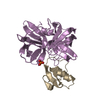
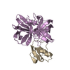
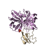
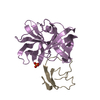
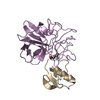
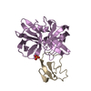
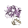
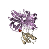
| Title | CRYSTAL STRUCTURE OF THE OMTKY3 P1 VARIANT OMTKY3-SER18I IN COMPLEX WITH SGPB | ||||||
 Components Components |
| ||||||
 Keywords Keywords | HYDROLASE/HYDROLASE INHIBITOR / ENZYME-INHIBITOR COMPLEX / BETA-BRANCHED P1 RESIDUE / HYDROLASE-HYDROLASE INHIBITOR COMPLEX | ||||||
| Function / homology |  Function and homology information Function and homology informationstreptogrisin B / molecular function inhibitor activity / serine-type endopeptidase inhibitor activity / serine-type endopeptidase activity / proteolysis / extracellular region Similarity search - Function | ||||||
| Biological species |   Streptomyces griseus (bacteria) Streptomyces griseus (bacteria) | ||||||
| Method |  X-RAY DIFFRACTION / Resolution: 1.8 Å X-RAY DIFFRACTION / Resolution: 1.8 Å | ||||||
 Authors Authors | Bateman, K.S. / Anderson, S. / Lu, W. / Qasim, M.A. / Laskowski Jr., M. / James, M.N.G. | ||||||
 Citation Citation | Journal: Protein Sci. / Year: 2000 Title: Deleterious effects of beta-branched residues in the S1 specificity pocket of Streptomyces griseus proteinase B (SGPB): crystal structures of the turkey ovomucoid third domain variants Ile18I, ...Title: Deleterious effects of beta-branched residues in the S1 specificity pocket of Streptomyces griseus proteinase B (SGPB): crystal structures of the turkey ovomucoid third domain variants Ile18I, Val18I, Thr18I, and Ser18I in complex with SGPB. Authors: Bateman, K.S. / Anderson, S. / Lu, W. / Qasim, M.A. / Laskowski Jr., M. / James, M.N. | ||||||
| History |
|
- Structure visualization
| Structure viewer | Molecule: MolmilJmol/JSmol |
|---|
- Downloads & links
-Download
| PDBx/mmCIF format | 1ct0.cif.gz | 58.8 KB | Display | PDBx/mmCIF format |
|---|---|---|---|---|
| PDB format | pdb1ct0.ent.gz | 41.4 KB | Display | PDB format |
| PDBx/mmJSON format | 1ct0.json.gz | Tree view | PDBx/mmJSON format | |
| Others |  Other downloads Other downloads |
-Validation report
| Summary document | 1ct0_validation.pdf.gz | 413.8 KB | Display | wwPDB validaton report |
|---|---|---|---|---|
| Full document | 1ct0_full_validation.pdf.gz | 415.1 KB | Display | |
| Data in XML | 1ct0_validation.xml.gz | 13.6 KB | Display | |
| Data in CIF | 1ct0_validation.cif.gz | 18.4 KB | Display | |
| Arichive directory | https://data.pdbj.org/pub/pdb/validation_reports/ct/1ct0ftp://data.pdbj.org/pub/pdb/validation_reports/ct/1ct0 | HTTPS FTP |
-Related structure data


-Links
 PDBj
PDBj
- Assembly
| Deposited unit | 
| ||||||||
|---|---|---|---|---|---|---|---|---|---|
| 1 |
| ||||||||
| Unit cell |
|
-Components
| #1: Protein | Mass: 18665.285 Da / Num. of mol.: 1 / Source method: isolated from a natural source / Source: (natural) Streptomyces griseus (bacteria) / Strain: K1 / References: UniProt: P00777, streptogrisin B |
|---|---|
| #2: Protein | Mass: 5559.208 Da / Num. of mol.: 1 / Fragment: THIRD DOMAIN SER18-OMTKY3 / Mutation: DEL 1-5, L18S Source method: isolated from a genetically manipulated source Source: (gene. exp.) |
| #3: Water | ChemComp-HOH /  Mass: 18.015 Da / Num. of mol.: 152 / Source method: isolated from a natural source / Formula: H2O Mass: 18.015 Da / Num. of mol.: 152 / Source method: isolated from a natural source / Formula: H2O |
| Has protein modification | Y |
-Experimental details
-Experiment
| Experiment | Method: X-RAY DIFFRACTION / Number of used crystals: 1 |
|---|
- Sample preparation
| Crystal | Density Matthews: 2.04 Å3/Da / Density % sol: 39.74 % | |||||||||||||||
|---|---|---|---|---|---|---|---|---|---|---|---|---|---|---|---|---|
| Crystal grow | Temperature: 298 K / Method: vapor diffusion, hanging drop / pH: 7.4 Details: PEG 4000 sodium potassium phosphate, pH 7.4, VAPOR DIFFUSION, HANGING DROP, temperature 298.0K | |||||||||||||||
| Crystal grow | *PLUS | |||||||||||||||
| Components of the solutions | *PLUS
|
-Data collection
| Diffraction | Mean temperature: 298 K |
|---|---|
| Diffraction source | Source: ROTATING ANODE / Type: RIGAKU / Wavelength: 1.5418 |
| Detector | Type: MAC Science DIP-2000H / Detector: IMAGE PLATE / Date: Sep 2, 1997 |
| Radiation | Protocol: SINGLE WAVELENGTH / Monochromatic (M) / Laue (L): M / Scattering type: x-ray |
| Radiation wavelength | Wavelength: 1.5418 Å / Relative weight: 1 |
| Reflection | Resolution: 1.8→20 Å / Num. all: 112593 / % possible obs: 96.7 % / Redundancy: 3.1 % / Biso Wilson estimate: 10.8 Å2 / Rmerge(I) obs: 0.122 / Net I/σ(I): 7.71 |
| Reflection shell | Resolution: 1.8→1.83 Å / Rmerge(I) obs: 0.455 / % possible all: 72 |
| Reflection | *PLUS Highest resolution: 1.9 Å / Num. obs: 15363 / % possible obs: 99.2 % / Num. measured all: 57863 / Rmerge(I) obs: 0.118 |
| Reflection shell | *PLUS Highest resolution: 1.9 Å / Lowest resolution: 1.93 Å / % possible obs: 99.4 % / Rmerge(I) obs: 0.33 / Mean I/σ(I) obs: 2.16 |
- Processing
| Software |
| |||||||||||||||||||||
|---|---|---|---|---|---|---|---|---|---|---|---|---|---|---|---|---|---|---|---|---|---|---|
| Refinement | Resolution: 1.8→20 Å / Stereochemistry target values: Engh & Huber
| |||||||||||||||||||||
| Refinement step | Cycle: LAST / Resolution: 1.8→20 Å
| |||||||||||||||||||||
| Refine LS restraints |
| |||||||||||||||||||||
| Software | *PLUS Name: TNT / Classification: refinement | |||||||||||||||||||||
| Refinement | *PLUS Highest resolution: 1.9 Å / Num. reflection all: 15363 / Rfactor all: 0.164 | |||||||||||||||||||||
| Solvent computation | *PLUS | |||||||||||||||||||||
| Displacement parameters | *PLUS | |||||||||||||||||||||
| Refine LS restraints | *PLUS Type: t_plane_restr / Dev ideal: 0.013 |
