 Movie
Movie Controller


+ Open data

- Basic information
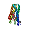
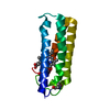
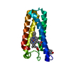
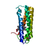
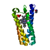
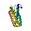
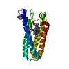
| Entry | Database: PDB / ID: 1cq4 | ||||||
|---|---|---|---|---|---|---|---|
| Title | CI2 MUTANT WITH TETRAGLUTAMINE (MGQQQQGM) REPLACING MET59 | ||||||
 Components Components | (PROTEIN (SERINE PROTEINASE INHIBITOR 2)) x 2 | ||||||
 Keywords Keywords | HYDROLASE INHIBITOR / SERINE PROTEASE INHIBITOR / POLYGLUTAMINE INSERTION MUTANT / SUBTILISIN- CHYMOTRYPSIN INHIBITOR-2 / IMMUNE SYSTEM | ||||||
| Function / homology | Proteinase inhibitor I13, potato inhibitor I / Proteinase inhibitor I13, potato inhibitor I superfamily / Potato inhibitor I family / Potato inhibitor I family signature. / serine-type endopeptidase inhibitor activity / response to wounding / Subtilisin-chymotrypsin inhibitor-2A Function and homology information Function and homology information | ||||||
| Biological species |  | ||||||
| Method |  X-RAY DIFFRACTION / SYNCHROTRON / MOLECULAR REPLACEMENT / Resolution: 1.8 Å X-RAY DIFFRACTION / SYNCHROTRON / MOLECULAR REPLACEMENT / Resolution: 1.8 Å | ||||||
 Authors Authors | Chen, Y.W. / Stott, K.R. | ||||||
 Citation Citation | Journal: Proc.Natl.Acad.Sci.USA / Year: 1999 Title: Crystal structure of a dimeric chymotrypsin inhibitor 2 mutant containing an inserted glutamine repeat. Authors: Chen, Y.W. / Stott, K. / Perutz, M.F. #1: Journal: J.Mol.Biol. / Year: 1997Title: Glutamine, Alanine or Glycine Repeats Inserted Into the Loop of a Protein Have Minimal Effects on Stability and Folding Rates Authors: Ladurner, A.G. / Fersht, A.R. #2: Journal: Fold.Des. / Year: 1996Title: Towards the Complete Structural Characterization of a Protein Folding Pathway: The Structures of the Denatured, Transition and Native States for the Association/Folding of Two Complementary ...Title: Towards the Complete Structural Characterization of a Protein Folding Pathway: The Structures of the Denatured, Transition and Native States for the Association/Folding of Two Complementary Fragments of Cleaved Chymotrypsin Inhibitor 2. Direction Evidence for a Nucleation-Condensation Mechanism Authors: Neira, J.L. / Davis, B. / Ladurner, A.G. / Buckle, A.M. / De Prat Gay, G. / Fersht, A.R. #3: Journal: Proc.Natl.Acad.Sci.USA / Year: 1995Title: Incorporation of Glutamine Repeats Makes Protein Oligomerize: Implications for Neurodegenerative Diseases Authors: Stott, K. / Blackburn, J.M. / Butler, P.J.G. / Perutz, M. #4: Journal: Biochemistry / Year: 1987Title: Crystal and Molecular Structure of the Serine Proteinase Inhibitor Ci-2 from Barley Seeds Authors: Mcphalen, C.A. / James, M.N.G. #5: Journal: Protein Eng. / Year: 1987Title: The Determination of the Three-Dimensional Structure of Barley Serine Proteinase Inhibitor 2 by Nuclear Magnetic Resonance, Distance Geometry and Restrained Molecular Dynamics Authors: Clore, G.M. / Gronenborn, A.M. / Kjaer, M. / Poulsen, F.M. | ||||||
| History |
|
- Structure visualization
| Structure viewer | Molecule: MolmilJmol/JSmol |
|---|
- Downloads & links
-Download
| PDBx/mmCIF format | 1cq4.cif.gz | 27.5 KB | Display | PDBx/mmCIF format |
|---|---|---|---|---|
| PDB format | pdb1cq4.ent.gz | 17.6 KB | Display | PDB format |
| PDBx/mmJSON format | 1cq4.json.gz | Tree view | PDBx/mmJSON format | |
| Others |  Other downloads Other downloads |
-Validation report
| Arichive directory | https://data.pdbj.org/pub/pdb/validation_reports/cq/1cq4ftp://data.pdbj.org/pub/pdb/validation_reports/cq/1cq4 | HTTPS FTP |
|---|
-Related structure data
| Related structure data |  2ci2S S: Starting model for refinement |
|---|---|
| Similar structure data |


-Links
 PDBj
PDBj- Assembly
| Deposited unit | 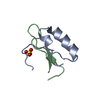
| ||||||||
|---|---|---|---|---|---|---|---|---|---|
| 1 | 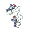
| ||||||||
| 2 | x 6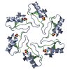
| ||||||||
| 3 | x 12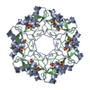
| ||||||||
| Unit cell |
|
-Components
| #1: Protein/peptide | Mass: 5212.127 Da / Num. of mol.: 1 / Mutation: YES Source method: isolated from a genetically manipulated source Details: EXISTS AS A1B2/A2B1 DOMAIN-SWAPPED DIMER / Source: (gene. exp.)  |
|---|---|
| #2: Protein/peptide | Mass: 2877.325 Da / Num. of mol.: 1 / Mutation: YES Source method: isolated from a genetically manipulated source Details: EXISTS AS A1B2/A2B1 DOMAIN-SWAPPED DIMER / Source: (gene. exp.) |
| #3: Chemical | ChemComp-SO4 /   Mass: 96.063 Da / Num. of mol.: 1 / Source method: obtained synthetically / Formula: SO4 Mass: 96.063 Da / Num. of mol.: 1 / Source method: obtained synthetically / Formula: SO4 |
| #4: Water | ChemComp-HOH /  Mass: 18.015 Da / Num. of mol.: 55 / Source method: isolated from a natural source / Formula: H2O Mass: 18.015 Da / Num. of mol.: 55 / Source method: isolated from a natural source / Formula: H2O |
-Experimental details
-Experiment
| Experiment | Method: X-RAY DIFFRACTION / Number of used crystals: 1 |
|---|
- Sample preparation
| Crystal | Density Matthews: 2.6 Å3/Da / Density % sol: 53 % | ||||||||||||||||||||||||||||||
|---|---|---|---|---|---|---|---|---|---|---|---|---|---|---|---|---|---|---|---|---|---|---|---|---|---|---|---|---|---|---|---|
| Crystal grow | pH: 7.5 Details: DROPS WERE PREPARED BY MIXING 2 MICROLITERS OF PURIFIED DIMER AT 25MG/ML WITH AN EQUAL VOLUME OF BUFFER (30% W/V PEG-400, 1.0 M LITHIUM SULPHATE, AND 1 MM CALCIUM CHLORIDE, IN 0.1 M TRIS-HCL ...Details: DROPS WERE PREPARED BY MIXING 2 MICROLITERS OF PURIFIED DIMER AT 25MG/ML WITH AN EQUAL VOLUME OF BUFFER (30% W/V PEG-400, 1.0 M LITHIUM SULPHATE, AND 1 MM CALCIUM CHLORIDE, IN 0.1 M TRIS-HCL AT PH 7.5), WHICH WAS ALSO USED AS THE WELL BUFFER (1 ML). | ||||||||||||||||||||||||||||||
| Crystal grow | *PLUS Temperature: 4 ℃ / Method: vapor diffusion, hanging drop | ||||||||||||||||||||||||||||||
| Components of the solutions | *PLUS
|
-Data collection
| Diffraction | Mean temperature: 100 K |
|---|---|
| Diffraction source | Source: SYNCHROTRON / Site: EMBL/DESY, HAMBURG  / Beamline: X11 / Wavelength: 0.912 / Beamline: X11 / Wavelength: 0.912 |
| Detector | Type: MARRESEARCH / Detector: IMAGE PLATE / Date: Jun 24, 1996 / Details: BENT MIRROR |
| Radiation | Protocol: SINGLE WAVELENGTH / Monochromatic (M) / Laue (L): M / Scattering type: x-ray |
| Radiation wavelength | Wavelength: 0.912 Å / Relative weight: 1 |
| Reflection | Resolution: 1.8→22.3 Å / Num. obs: 8153 / % possible obs: 99.7 % / Redundancy: 10.6 % / Biso Wilson estimate: 23.5 Å2 / Rmerge(I) obs: 0.081 / Rsym value: 0.078 / Net I/σ(I): 3.6 |
| Reflection shell | Resolution: 1.8→1.9 Å / Redundancy: 10.8 % / Rmerge(I) obs: 0.338 / Mean I/σ(I) obs: 2.3 / Rsym value: 0.325 / % possible all: 100 |
| Reflection | *PLUS Num. measured all: 86441 / Rmerge(I) obs: 0.074 |
| Reflection shell | *PLUS % possible obs: 100 % / Rmerge(I) obs: 0.286 |
- Processing
| Software |
| ||||||||||||||||||||||||||||||||||||||||||||||||||||||||||||||||||||||||||||||||||||
|---|---|---|---|---|---|---|---|---|---|---|---|---|---|---|---|---|---|---|---|---|---|---|---|---|---|---|---|---|---|---|---|---|---|---|---|---|---|---|---|---|---|---|---|---|---|---|---|---|---|---|---|---|---|---|---|---|---|---|---|---|---|---|---|---|---|---|---|---|---|---|---|---|---|---|---|---|---|---|---|---|---|---|---|---|---|
| Refinement | Method to determine structure: MOLECULAR REPLACEMENT Starting model: PDB ENTRY 2CI2 Resolution: 1.8→22 Å / SU B: 2.42 / SU ML: 0.08 / Cross valid method: THROUGHOUT / σ(F): 0 / ESU R: 0.14 / ESU R Free: 0.15
| ||||||||||||||||||||||||||||||||||||||||||||||||||||||||||||||||||||||||||||||||||||
| Displacement parameters | Biso mean: 32.2 Å2
| ||||||||||||||||||||||||||||||||||||||||||||||||||||||||||||||||||||||||||||||||||||
| Refinement step | Cycle: LAST / Resolution: 1.8→22 Å
| ||||||||||||||||||||||||||||||||||||||||||||||||||||||||||||||||||||||||||||||||||||
| Refine LS restraints |
|
