 Movie
Movie Controller

[English] 日本語
 Yorodumi
- PDB-1col: REFINED STRUCTURE OF THE PORE-FORMING DOMAIN OF COLICIN A AT 2.4 ... -
+ Open data

- Basic information
| Entry | Database: PDB / ID: 1col | ||||||
|---|---|---|---|---|---|---|---|
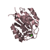
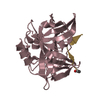
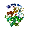
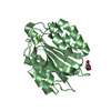
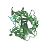
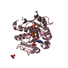
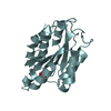
| Title | REFINED STRUCTURE OF THE PORE-FORMING DOMAIN OF COLICIN A AT 2.4 ANGSTROMS RESOLUTION | ||||||
 Components Components | COLICIN A | ||||||
 Keywords Keywords | ANTIBACTERIAL PROTEIN | ||||||
| Function / homology |  Function and homology information Function and homology informationpore-forming activity / killing of cells of another organism / defense response to Gram-negative bacterium / plasma membrane Similarity search - Function | ||||||
| Biological species |  | ||||||
| Method |  X-RAY DIFFRACTION / Resolution: 2.4 Å X-RAY DIFFRACTION / Resolution: 2.4 Å | ||||||
 Authors Authors | Parker, M.W. / Postma, J.P.M. / Pattus, F. / Tucker, A.D. / Tsernoglou, D. | ||||||
 Citation Citation | Journal: J.Mol.Biol. / Year: 1992 Title: Refined structure of the pore-forming domain of colicin A at 2.4 A resolution. Authors: Parker, M.W. / Postma, J.P. / Pattus, F. / Tucker, A.D. / Tsernoglou, D. #1: Journal: J.Mol.Biol. / Year: 1991Title: Fluorescence Energy Transfer Distance Measurements Using Site-Directed Single Cysteine Mutants Authors: Lakey, J.H. / Baty, D. / Pattus, F. #2: Journal: Experientia / Year: 1990Title: Colicins: Prokaryotic Killer-Pores Authors: Pattus, F. / Massotte, D. / Wilmsen, H.U. / Lakey, J. / Tsernoglou, D. / Tucker, A. / Parker, M.W. #3: Journal: Nature / Year: 1989Title: Structure of the Membrane-Pore-Forming Fragment of Colicin A Authors: Parker, M.W. / Pattus, F. / Tucker, A.D. / Tsernoglou, D. #4: Journal: Biochim.Biophys.Acta / Year: 1988Title: The Membrane Channel-Forming Colicin A: Synthesis, Secretion, Structure, Action and Immunity Authors: Lazdunski, C.J. / Baty, D. / Geli, V. / Cavard, D. / Morlon, J. / Howard, R.Lloubes.S.P. / Knibiehler, M. / Chartier, M. / Varenne, S. / Frenette, M. / Dasseux, J.-L. / Pattus, F. #5: Journal: Eur.Biophys.J. / Year: 1987Title: Gating Processes of Channels Induced by Colicin A, its C-Terminal Fragment and Colicin E1 in Planar Lipid Bilayers Authors: Collarini, M. / Amblard, G. / Lazdunski, C. / Pattus, F. #6: Journal: J.Mol.Biol. / Year: 1986Title: Crystallization of the C-Terminal Domain of Colicin A Carrying the Voltage-Dependent Pore Activity of the Protein Authors: Tucker, A.D. / Pattus, F. / Tsernoglou, D. | ||||||
| History |
|
- Structure visualization
| Structure viewer | Molecule: MolmilJmol/JSmol |
|---|
- Downloads & links
-Download
| PDBx/mmCIF format | 1col.cif.gz | 84.4 KB | Display | PDBx/mmCIF format |
|---|---|---|---|---|
| PDB format | pdb1col.ent.gz | 65.6 KB | Display | PDB format |
| PDBx/mmJSON format | 1col.json.gz | Tree view | PDBx/mmJSON format | |
| Others |  Other downloads Other downloads |
-Validation report
| Arichive directory | https://data.pdbj.org/pub/pdb/validation_reports/co/1colftp://data.pdbj.org/pub/pdb/validation_reports/co/1col | HTTPS FTP |
|---|
-Related structure data
| Similar structure data |
|---|


-Links
 PDBj
PDBj- Assembly
| Deposited unit | 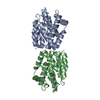
| ||||||||
|---|---|---|---|---|---|---|---|---|---|
| 1 | 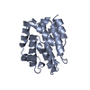
| ||||||||
| 2 | 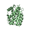
| ||||||||
| Unit cell |
| ||||||||
| Noncrystallographic symmetry (NCS) | NCS oper: (Code: given Matrix: (0.47345, -0.01458, 0.88043), Vector: |
-Components
| #1: Protein | Mass: 21818.148 Da / Num. of mol.: 2 Source method: isolated from a genetically manipulated source Source: (gene. exp.) #2: Water | ChemComp-HOH / |  Mass: 18.015 Da / Num. of mol.: 83 / Source method: isolated from a natural source / Formula: H2O Mass: 18.015 Da / Num. of mol.: 83 / Source method: isolated from a natural source / Formula: H2O |
|---|
-Experimental details
-Experiment
| Experiment | Method: X-RAY DIFFRACTION |
|---|
- Sample preparation
| Crystal | Density Matthews: 2.62 Å3/Da / Density % sol: 53.04 % | ||||||||||||||||||||||||||||||||||||
|---|---|---|---|---|---|---|---|---|---|---|---|---|---|---|---|---|---|---|---|---|---|---|---|---|---|---|---|---|---|---|---|---|---|---|---|---|---|
| Crystal grow | *PLUS Method: vapor diffusion, hanging drop / Details: referred to J.Mol.Biol. 190.133-134 1986 | ||||||||||||||||||||||||||||||||||||
| Components of the solutions | *PLUS
|
-Data collection
| Radiation | Scattering type: x-ray |
|---|---|
| Radiation wavelength | Relative weight: 1 |
| Reflection | *PLUS Highest resolution: 2.5 Å / Lowest resolution: 6 Å / Num. obs: 10024 / Observed criterion σ(F): 3 / Rmerge F obs: 0.244 |
- Processing
| Software | Name: PROLSQ / Classification: refinement | ||||||||||||||||||||||||||||||||||||||||||||||||||||||||||||||||||||||||||||||||||||
|---|---|---|---|---|---|---|---|---|---|---|---|---|---|---|---|---|---|---|---|---|---|---|---|---|---|---|---|---|---|---|---|---|---|---|---|---|---|---|---|---|---|---|---|---|---|---|---|---|---|---|---|---|---|---|---|---|---|---|---|---|---|---|---|---|---|---|---|---|---|---|---|---|---|---|---|---|---|---|---|---|---|---|---|---|---|
| Refinement | Resolution: 2.4→6 Å / Rfactor obs: 0.18 | ||||||||||||||||||||||||||||||||||||||||||||||||||||||||||||||||||||||||||||||||||||
| Refinement step | Cycle: LAST / Resolution: 2.4→6 Å
| ||||||||||||||||||||||||||||||||||||||||||||||||||||||||||||||||||||||||||||||||||||
| Refine LS restraints |
| ||||||||||||||||||||||||||||||||||||||||||||||||||||||||||||||||||||||||||||||||||||
| Refinement | *PLUS Highest resolution: 2.5 Å / Lowest resolution: 6 Å / σ(F): 2 / Rfactor obs: 0.26 | ||||||||||||||||||||||||||||||||||||||||||||||||||||||||||||||||||||||||||||||||||||
| Solvent computation | *PLUS | ||||||||||||||||||||||||||||||||||||||||||||||||||||||||||||||||||||||||||||||||||||
| Displacement parameters | *PLUS |
