 Movie
Movie Controller

[English] 日本語
 Yorodumi
- PDB-1clk: CRYSTAL STRUCTURE OF STREPTOMYCES DIASTATICUS NO.7 STRAIN M1033 X... -
+ Open data

- Basic information
| Entry | Database: PDB / ID: 1clk | ||||||
|---|---|---|---|---|---|---|---|
| Title | CRYSTAL STRUCTURE OF STREPTOMYCES DIASTATICUS NO.7 STRAIN M1033 XYLOSE ISOMERASE AT 1.9 A RESOLUTION WITH PSEUDO-I222 SPACE GROUP | ||||||
 Components Components | XYLOSE ISOMERASE | ||||||
 Keywords Keywords | ISOMERASE / XYLOSE ISOMERASE / GLUCOSE ISOMERASE / STREPTOMYCES / PSEUDO-I222 | ||||||
| Function / homology |  Function and homology information Function and homology informationxylose isomerase / xylose isomerase activity / D-xylose metabolic process / magnesium ion binding / cytoplasm Similarity search - Function | ||||||
| Biological species |   Streptomyces diastaticus (bacteria) Streptomyces diastaticus (bacteria) | ||||||
| Method |  X-RAY DIFFRACTION / MOLECULAR REPLACEMENT / Resolution: 1.9 Å X-RAY DIFFRACTION / MOLECULAR REPLACEMENT / Resolution: 1.9 Å | ||||||
 Authors Authors | Niu, L. / Teng, M. / Zhu, X. / Gong, W. | ||||||
 Citation Citation | Journal: Acta Crystallogr.,Sect.D / Year: 2000 Title: Structure of xylose isomerase from Streptomyces diastaticus no. 7 strain M1033 at 1.85 A resolution. Authors: Zhu, X. / Teng, M. / Niu, L. / Xu, C. / Wang, Y. #1: Journal: SCI.CHINA, SER.C: LIFE SCI. / Year: 1996Title: Crystal Structure of Streptomyces diastaticus No.7 Strain M1033 Xylose Isomerase Authors: Zhu, X. / Gong, W. / Niu, L. / Teng, M. / Xu, C. / Wu, C. / Cui, T. / Wang, Y. / Wang, C. #2: Journal: Chin.Sci.Bull. / Year: 1991Title: Growth of Single Crystals and Preliminary Analysis of Glucose Isomerase From Streptomyces M1033 Authors: Zhang, G.Y. / Niu, L.W. / Huang, W.Z. / Wang, C. / Liu, J. / Cui, Y. / Liu, X.A. / Wang, Y.Z. / Xu, X. / Liang, D.C. | ||||||
| History |
|
- Structure visualization
| Structure viewer | Molecule: MolmilJmol/JSmol |
|---|
- Downloads & links
-Download
| PDBx/mmCIF format | 1clk.cif.gz | 98.1 KB | Display | PDBx/mmCIF format |
|---|---|---|---|---|
| PDB format | pdb1clk.ent.gz | 72 KB | Display | PDB format |
| PDBx/mmJSON format | 1clk.json.gz | Tree view | PDBx/mmJSON format | |
| Others |  Other downloads Other downloads |
-Validation report
| Arichive directory | https://data.pdbj.org/pub/pdb/validation_reports/cl/1clkftp://data.pdbj.org/pub/pdb/validation_reports/cl/1clk | HTTPS FTP |
|---|
-Related structure data
| Related structure data |  1qt1C  7xia S: Starting model for refinement C: citing same article ( |
|---|---|
| Similar structure data |

-Links
 PDBj
PDBj

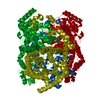
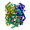
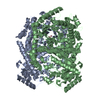
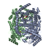
- Assembly
| Deposited unit | 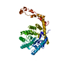
| ||||||||
|---|---|---|---|---|---|---|---|---|---|
| 1 | 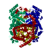
| ||||||||
| Unit cell |
|
-Components
| #1: Protein | Mass: 42709.664 Da / Num. of mol.: 1 / Source method: isolated from a natural source / Source: (natural) Streptomyces diastaticus (bacteria) / Strain: STREPTOMYCES DIASTATICUS NO.7 STRAIN M1033 / References: UniProt: P50910, xylose isomerase |
|---|---|
| #2: Chemical | ChemComp-MG /   Mass: 24.305 Da / Num. of mol.: 1 / Source method: obtained synthetically / Formula: Mg Mass: 24.305 Da / Num. of mol.: 1 / Source method: obtained synthetically / Formula: Mg |
| #3: Chemical | ChemComp-CO /   Mass: 58.933 Da / Num. of mol.: 1 / Source method: obtained synthetically / Formula: Co Mass: 58.933 Da / Num. of mol.: 1 / Source method: obtained synthetically / Formula: Co |
| #4: Water | ChemComp-HOH /  Mass: 18.015 Da / Num. of mol.: 392 / Source method: isolated from a natural source / Formula: H2O Mass: 18.015 Da / Num. of mol.: 392 / Source method: isolated from a natural source / Formula: H2O |
-Experimental details
-Experiment
| Experiment | Method: X-RAY DIFFRACTION / Number of used crystals: 1 |
|---|
- Sample preparation
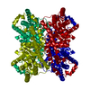
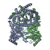
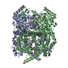
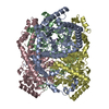
| Crystal | Density Matthews: 2.39 Å3/Da / Density % sol: 48.51 % Description: THE STRUCTURE HAS BEEN SOLVED IN PSEUDO-I222 SPACE GROUP. THE MOLECULE IS A TETRAMER CONTAINING ONE SUBUNITS IN THE ASYMMETRIC UNIT. TETRAMERS ARE POSITIONED ON THE 222 SYMMETRY SITE AT ...Description: THE STRUCTURE HAS BEEN SOLVED IN PSEUDO-I222 SPACE GROUP. THE MOLECULE IS A TETRAMER CONTAINING ONE SUBUNITS IN THE ASYMMETRIC UNIT. TETRAMERS ARE POSITIONED ON THE 222 SYMMETRY SITE AT THE ORIGIN OF THE CELL. THE STRUCTURE OF THE MONOMER IS AN EIGHT-FOLD ALPHA-BETA BARREL WITH AN EXTENDED C-TERMINAL LOOP WHICH FACILITATES AGGREGATION OF MONOMERS TO TETRAMERS. |
|---|---|
| Crystal grow | pH: 7.5 / Details: pH 7.5 |
-Data collection
| Diffraction | Mean temperature: 287 K |
|---|---|
| Diffraction source | Source: ROTATING ANODE / Type: RIGAKU / Wavelength: 1.5418 |
| Detector | Type: SIEMENS / Detector: AREA DETECTOR |
| Radiation | Monochromator: NI FILTER / Protocol: SINGLE WAVELENGTH / Monochromatic (M) / Laue (L): M / Scattering type: x-ray |
| Radiation wavelength | Wavelength: 1.5418 Å / Relative weight: 1 |
| Reflection | Resolution: 1.9→5 Å / Num. obs: 25700 / % possible obs: 83.74 % / Redundancy: 2 % / Rmerge(I) obs: 0.085 / Net I/σ(I): 11.52 |
| Reflection shell | Resolution: 1.9→1.98 Å / Mean I/σ(I) obs: 4.03 / % possible all: 75.56 |
- Processing
| Software |
| ||||||||||||||||||||||||||||||||||||||||||||||||||||||||||||
|---|---|---|---|---|---|---|---|---|---|---|---|---|---|---|---|---|---|---|---|---|---|---|---|---|---|---|---|---|---|---|---|---|---|---|---|---|---|---|---|---|---|---|---|---|---|---|---|---|---|---|---|---|---|---|---|---|---|---|---|---|---|
| Refinement | Method to determine structure: MOLECULAR REPLACEMENT Starting model: PDB ENTRY 7XIA 7xia Resolution: 1.9→5 Å / Data cutoff low absF: 5 / Isotropic thermal model: RESTRAINED / σ(F): 2
| ||||||||||||||||||||||||||||||||||||||||||||||||||||||||||||
| Displacement parameters | Biso mean: 19.95 Å2 | ||||||||||||||||||||||||||||||||||||||||||||||||||||||||||||
| Refine analyze | Luzzati coordinate error obs: 0.3 Å | ||||||||||||||||||||||||||||||||||||||||||||||||||||||||||||
| Refinement step | Cycle: LAST / Resolution: 1.9→5 Å
| ||||||||||||||||||||||||||||||||||||||||||||||||||||||||||||
| Refine LS restraints |
| ||||||||||||||||||||||||||||||||||||||||||||||||||||||||||||
| LS refinement shell | Resolution: 1.9→1.98 Å / Total num. of bins used: 8
| ||||||||||||||||||||||||||||||||||||||||||||||||||||||||||||
| Xplor file | Serial no: 1 / Param file: PARHCSDX.PRO / Topol file: TOPHCSDX.PRO |
