 Movie
Movie Controller

[English] 日本語
 Yorodumi
- PDB-1cf7: STRUCTURAL BASIS OF DNA RECOGNITION BY THE HETERODIMERIC CELL CYC... -
+ Open data

- Basic information
| Entry | Database: PDB / ID: 1cf7 | ||||||
|---|---|---|---|---|---|---|---|
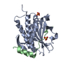
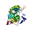
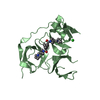
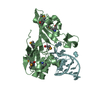
| Title | STRUCTURAL BASIS OF DNA RECOGNITION BY THE HETERODIMERIC CELL CYCLE TRANSCRIPTION FACTOR E2F-DP | ||||||
 Components Components |
| ||||||
 Keywords Keywords | TRANSCRIPTION/DNA / E2F / DP / WINGED-HELIX / DNA-BINDING DOMAIN / TRANSCRIPTION FACTOR / CELL CYCLE / TRANSCRIPTION-DNA COMPLEX | ||||||
| Function / homology |  Function and homology information Function and homology informationmulti-ciliated epithelial cell differentiation / centriole assembly / motile cilium assembly / Inhibition of replication initiation of damaged DNA by RB1/E2F1 / Transcription of E2F targets under negative control by p107 (RBL1) and p130 (RBL2) in complex with HDAC1 / Transcription of E2F targets under negative control by DREAM complex / Activation of NOXA and translocation to mitochondria / Activation of PUMA and translocation to mitochondria / transcription factor binding / G1/S-Specific Transcription ...multi-ciliated epithelial cell differentiation / centriole assembly / motile cilium assembly / Inhibition of replication initiation of damaged DNA by RB1/E2F1 / Transcription of E2F targets under negative control by p107 (RBL1) and p130 (RBL2) in complex with HDAC1 / Transcription of E2F targets under negative control by DREAM complex / Activation of NOXA and translocation to mitochondria / Activation of PUMA and translocation to mitochondria / transcription factor binding / G1/S-Specific Transcription / Defective binding of RB1 mutants to E2F1,(E2F2, E2F3) / Transcriptional Regulation by E2F6 / G0 and Early G1 / Cyclin E associated events during G1/S transition / Cyclin A:Cdk2-associated events at S phase entry / TP53 Regulates Transcription of Genes Involved in G2 Cell Cycle Arrest / promoter-specific chromatin binding / SMAD2/SMAD3:SMAD4 heterotrimer regulates transcription / Oncogene Induced Senescence / Pre-NOTCH Transcription and Translation / RNA polymerase II transcription regulator complex / Transcriptional regulation of granulopoiesis / sequence-specific double-stranded DNA binding / Cyclin D associated events in G1 / mitotic spindle / DNA-binding transcription activator activity, RNA polymerase II-specific / Oxidative Stress Induced Senescence / DNA-binding transcription factor activity, RNA polymerase II-specific / protein dimerization activity / regulation of cell cycle / RNA polymerase II cis-regulatory region sequence-specific DNA binding / DNA-binding transcription factor activity / protein domain specific binding / negative regulation of DNA-templated transcription / regulation of transcription by RNA polymerase II / regulation of DNA-templated transcription / chromatin / DNA-templated transcription / positive regulation of transcription by RNA polymerase II / DNA binding / nucleoplasm / nucleus / cytoplasm Similarity search - Function | ||||||
| Biological species |  Homo sapiens (human) Homo sapiens (human) | ||||||
| Method |  X-RAY DIFFRACTION / SYNCHROTRON / MIR / Resolution: 2.6 Å X-RAY DIFFRACTION / SYNCHROTRON / MIR / Resolution: 2.6 Å | ||||||
 Authors Authors | Zheng, N. / Fraenkel, E. / Pabo, C.O. / Pavletich, N.P. | ||||||
 Citation Citation | Journal: Genes Dev. / Year: 1999 Title: Structural basis of DNA recognition by the heterodimeric cell cycle transcription factor E2F-DP. Authors: Zheng, N. / Fraenkel, E. / Pabo, C.O. / Pavletich, N.P. | ||||||
| History |
|
- Structure visualization
| Structure viewer | Molecule: MolmilJmol/JSmol |
|---|
- Downloads & links
-Download
| PDBx/mmCIF format | 1cf7.cif.gz | 61.2 KB | Display | PDBx/mmCIF format |
|---|---|---|---|---|
| PDB format | pdb1cf7.ent.gz | 42.4 KB | Display | PDB format |
| PDBx/mmJSON format | 1cf7.json.gz | Tree view | PDBx/mmJSON format | |
| Others |  Other downloads Other downloads |
-Validation report
| Arichive directory | https://data.pdbj.org/pub/pdb/validation_reports/cf/1cf7ftp://data.pdbj.org/pub/pdb/validation_reports/cf/1cf7 | HTTPS FTP |
|---|
-Related structure data
| Similar structure data |
|---|


-Links
 PDBj
PDBj


- Assembly
| Deposited unit | 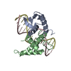
| ||||||||||
|---|---|---|---|---|---|---|---|---|---|---|---|
| 1 |
| ||||||||||
| Unit cell |
|
-Components
| #1: DNA chain | Mass: 4886.160 Da / Num. of mol.: 1 / Fragment: ADENOVIRUS TYPE 5 E2 PROMOTER E2F-BINDING SITE / Source method: obtained synthetically |
|---|---|
| #2: DNA chain | Mass: 4909.235 Da / Num. of mol.: 1 / Fragment: ADENOVIRUS TYPE 5 E2 PROMOTER E2F-BINDING SITE / Source method: obtained synthetically |
| #3: Protein | Mass: 8352.729 Da / Num. of mol.: 1 / Fragment: DNA-BINDING DOMAIN Source method: isolated from a genetically manipulated source Source: (gene. exp.) Homo sapiens (human) / Plasmid: PGEX-4T3 / Cell line (production host): BL21(DE3) / Cellular location (production host): CYTOPLASM / Production host:  |
| #4: Protein | Mass: 10836.347 Da / Num. of mol.: 1 / Fragment: DNA-BINDING DOMAIN Source method: isolated from a genetically manipulated source Source: (gene. exp.) Homo sapiens (human) / Plasmid: PGEX-4T3 / Cell line (production host): BL21(DE3) / Cellular location (production host): CYTOPLASM / Production host: |
| #5: Water | ChemComp-HOH /  Mass: 18.015 Da / Num. of mol.: 75 / Source method: isolated from a natural source / Formula: H2O Mass: 18.015 Da / Num. of mol.: 75 / Source method: isolated from a natural source / Formula: H2O |
-Experimental details
-Experiment
| Experiment | Method: X-RAY DIFFRACTION / Number of used crystals: 1 |
|---|
- Sample preparation
| Crystal | Density Matthews: 3.71 Å3/Da / Density % sol: 66.83 % | ||||||||||||||||||||||||||||||||||||||||||||||||||||||||||||||||||||||
|---|---|---|---|---|---|---|---|---|---|---|---|---|---|---|---|---|---|---|---|---|---|---|---|---|---|---|---|---|---|---|---|---|---|---|---|---|---|---|---|---|---|---|---|---|---|---|---|---|---|---|---|---|---|---|---|---|---|---|---|---|---|---|---|---|---|---|---|---|---|---|---|
| Crystal grow | Temperature: 277 K / pH: 6 Details: 14-16% PEG 400, 25MM NACL, 50MM MES, 10MM DTT, PH=6.0, 4 DEGREES C, temperature 277K | ||||||||||||||||||||||||||||||||||||||||||||||||||||||||||||||||||||||
| Components of the solutions |
| ||||||||||||||||||||||||||||||||||||||||||||||||||||||||||||||||||||||
| Crystal grow | *PLUS Temperature: 4 ℃ / Method: vapor diffusion, hanging drop | ||||||||||||||||||||||||||||||||||||||||||||||||||||||||||||||||||||||
| Components of the solutions | *PLUS
|
-Data collection
| Diffraction | Mean temperature: 127 K |
|---|---|
| Diffraction source | Source: SYNCHROTRON / Site: CHESS  / Beamline: A1 / Wavelength: 1.1548 / Beamline: A1 / Wavelength: 1.1548 |
| Radiation | Protocol: SINGLE WAVELENGTH / Monochromatic (M) / Laue (L): M / Scattering type: x-ray |
| Radiation wavelength | Wavelength: 1.1548 Å / Relative weight: 1 |
| Reflection | Resolution: 2.6→20 Å / Num. obs: 12901 / % possible obs: 96.5 % / Redundancy: 2.6 % / Rsym value: 5.4 |
| Reflection | *PLUS Num. measured all: 41452 / Rmerge(I) obs: 0.054 |
- Processing
| Software | Name: CNS / Version: 0.3A / Classification: refinement | ||||||||||||||||||||||||||||||||||||||||||||||||||||||||||||
|---|---|---|---|---|---|---|---|---|---|---|---|---|---|---|---|---|---|---|---|---|---|---|---|---|---|---|---|---|---|---|---|---|---|---|---|---|---|---|---|---|---|---|---|---|---|---|---|---|---|---|---|---|---|---|---|---|---|---|---|---|---|
| Refinement | Method to determine structure: MIR / Resolution: 2.6→15 Å / Cross valid method: THROUGHOUT / σ(F): 0
| ||||||||||||||||||||||||||||||||||||||||||||||||||||||||||||
| Refinement step | Cycle: LAST / Resolution: 2.6→15 Å
| ||||||||||||||||||||||||||||||||||||||||||||||||||||||||||||
| Refine LS restraints |
| ||||||||||||||||||||||||||||||||||||||||||||||||||||||||||||
| Software | *PLUS Version: 0.3A / Classification: refinement | ||||||||||||||||||||||||||||||||||||||||||||||||||||||||||||
| Refinement | *PLUS Highest resolution: 2.6 Å / Lowest resolution: 15 Å / σ(F): 0 / % reflection Rfree: 5 % / Rfactor obs: 0.229 / Rfactor Rfree: 0.283 | ||||||||||||||||||||||||||||||||||||||||||||||||||||||||||||
| Solvent computation | *PLUS | ||||||||||||||||||||||||||||||||||||||||||||||||||||||||||||
| Displacement parameters | *PLUS | ||||||||||||||||||||||||||||||||||||||||||||||||||||||||||||
| Refine LS restraints | *PLUS
|
