 Movie
Movie Controller

[English] 日本語
 Yorodumi
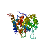
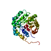
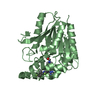
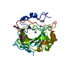
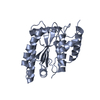
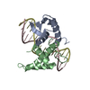
- PDB-1z97: Human Carbonic Anhydrase III: Structural and Kinetic Study of Cat... -
+ Open data

- Basic information
| Entry | Database: PDB / ID: 1z97 | ||||||
|---|---|---|---|---|---|---|---|
| Title | Human Carbonic Anhydrase III: Structural and Kinetic Study of Catalysis and Proton Transfer. | ||||||
 Components Components | Carbonic anhydrase III | ||||||
 Keywords Keywords | LYASE / carbonic anhydrase / proton wire / chemical rescue | ||||||
| Function / homology |  Function and homology information Function and homology informationcellular response to leptin stimulus / negative regulation of intrinsic apoptotic signaling pathway in response to hydrogen peroxide / response to lipid / nickel cation binding / phosphatase activity / negative regulation of reactive oxygen species biosynthetic process / skeletal muscle contraction / liver regeneration / Reversible hydration of carbon dioxide / carbonic anhydrase ...cellular response to leptin stimulus / negative regulation of intrinsic apoptotic signaling pathway in response to hydrogen peroxide / response to lipid / nickel cation binding / phosphatase activity / negative regulation of reactive oxygen species biosynthetic process / skeletal muscle contraction / liver regeneration / Reversible hydration of carbon dioxide / carbonic anhydrase / carbonate dehydratase activity / response to bacterium / sarcomere / cellular response to insulin stimulus / response to oxidative stress / cellular response to hypoxia / response to ethanol / positive regulation of cell population proliferation / zinc ion binding / nucleus / cytoplasm / cytosol Similarity search - Function | ||||||
| Biological species |  Homo sapiens (human) Homo sapiens (human) | ||||||
| Method |  X-RAY DIFFRACTION / MOLECULAR REPLACEMENT / Resolution: 2.1 Å X-RAY DIFFRACTION / MOLECULAR REPLACEMENT / Resolution: 2.1 Å | ||||||
 Authors Authors | Duda, D.M. / Tu, C. / Fisher, S.Z. / An, H. / Yoshioka, C. / Govindasamy, L. / Laipis, P.J. / Agbandje-McKenna, M. / Silverman, D.N. / McKenna, R. | ||||||
 Citation Citation | Journal: Biochemistry / Year: 2005 Title: Human Carbonic Anhydrase III: Structural and Kinetic Study of Catalysis and Proton Transfer Authors: Duda, D.M. / Tu, C. / Fisher, S.Z. / An, H. / Yoshioka, C. / Govindasamy, L. / Laipis, P.J. / Agbandje-McKenna, M. / Silverman, D.N. / McKenna, R. | ||||||
| History |
|
- Structure visualization
| Structure viewer | Molecule: MolmilJmol/JSmol |
|---|
- Downloads & links
-Download
| PDBx/mmCIF format | 1z97.cif.gz | 67.8 KB | Display | PDBx/mmCIF format |
|---|---|---|---|---|
| PDB format | pdb1z97.ent.gz | 48.9 KB | Display | PDB format |
| PDBx/mmJSON format | 1z97.json.gz | Tree view | PDBx/mmJSON format | |
| Others |  Other downloads Other downloads |
-Validation report
| Arichive directory | https://data.pdbj.org/pub/pdb/validation_reports/z9/1z97ftp://data.pdbj.org/pub/pdb/validation_reports/z9/1z97 | HTTPS FTP |
|---|
-Related structure data
| Related structure data |  1z93C  1fljS S: Starting model for refinement C: citing same article ( |
|---|---|
| Similar structure data |


-Links
 PDBj
PDBj
- Assembly
| Deposited unit | 
| ||||||||
|---|---|---|---|---|---|---|---|---|---|
| 1 |
| ||||||||
| Unit cell |
|
-Components
| #1: Protein | Mass: 30376.203 Da / Num. of mol.: 1 / Mutation: F198L, C182S, C187S Source method: isolated from a genetically manipulated source Source: (gene. exp.) Homo sapiens (human) / Gene: CA3 / Plasmid: pET-81f1 / Production host:  |
|---|---|
| #2: Chemical | ChemComp-ZN /   Mass: 65.409 Da / Num. of mol.: 1 / Source method: obtained synthetically / Formula: Zn Mass: 65.409 Da / Num. of mol.: 1 / Source method: obtained synthetically / Formula: Zn |
| #3: Water | ChemComp-HOH /  Mass: 18.015 Da / Num. of mol.: 97 / Source method: isolated from a natural source / Formula: H2O Mass: 18.015 Da / Num. of mol.: 97 / Source method: isolated from a natural source / Formula: H2O |
-Experimental details
-Experiment
| Experiment | Method: X-RAY DIFFRACTION / Number of used crystals: 1 |
|---|
- Sample preparation
| Crystal | Density Matthews: 2.07 Å3/Da / Density % sol: 40.55 % |
|---|---|
| Crystal grow | Temperature: 273 K / Method: vapor diffusion, hanging drop / pH: 9.2 Details: 30% PEG 8000, 10mM Tris, pH 9.2, VAPOR DIFFUSION, HANGING DROP, temperature 273K |
-Data collection
| Diffraction | Mean temperature: 293 K |
|---|---|
| Diffraction source | Source: ROTATING ANODE / Type: RIGAKU RU300 / Wavelength: 1.5418 Å |
| Detector | Type: RIGAKU RAXIS IV / Detector: IMAGE PLATE / Date: Jan 9, 2002 / Details: Osmic mirror |
| Radiation | Protocol: SINGLE WAVELENGTH / Monochromatic (M) / Laue (L): M / Scattering type: x-ray |
| Radiation wavelength | Wavelength: 1.5418 Å / Relative weight: 1 |
| Reflection | Resolution: 2.1→20 Å / Num. obs: 13525 / % possible obs: 92.3 % / Observed criterion σ(F): 3 / Rsym value: 0.081 |
| Reflection shell | Resolution: 2.1→2.17 Å / Num. unique all: 1268 / Rsym value: 0.341 / % possible all: 87.7 |
- Processing
| Software |
| |||||||||||||||||||||||||
|---|---|---|---|---|---|---|---|---|---|---|---|---|---|---|---|---|---|---|---|---|---|---|---|---|---|---|
| Refinement | Method to determine structure: MOLECULAR REPLACEMENT Starting model: pdb entry 1FLJ Resolution: 2.1→20 Å / Cross valid method: THROUGHOUT / Stereochemistry target values: Engh & Huber
| |||||||||||||||||||||||||
| Refinement step | Cycle: LAST / Resolution: 2.1→20 Å
| |||||||||||||||||||||||||
| Refine LS restraints |
|
