 Movie
Movie Controller


+ Open data

- Basic information
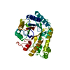
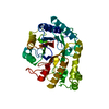
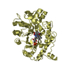
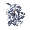
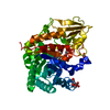
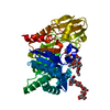
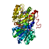
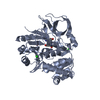
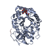
| Entry | Database: PDB / ID: 1ceo | ||||||
|---|---|---|---|---|---|---|---|
| Title | CELLULASE (CELC) MUTANT WITH GLU 140 REPLACED BY GLN | ||||||
 Components Components | CELLULASE CELC | ||||||
 Keywords Keywords | CELLULOSE DEGRADATION / GLYCOSYL HYDROLASE / CELLULASE / FAMILY A/5 OF GLYCOSYL HYDROLASES / CLOSTRIDIUM THERMOCELLUM | ||||||
| Function / homology |  Function and homology information Function and homology informationcellulase / cellulase activity / beta-glucosidase activity / cellulose catabolic process / cell surface / extracellular region Similarity search - Function | ||||||
| Biological species |  Clostridium thermocellum (bacteria) Clostridium thermocellum (bacteria) | ||||||
| Method |  X-RAY DIFFRACTION / SYNCHROTRON / Resolution: 1.9 Å X-RAY DIFFRACTION / SYNCHROTRON / Resolution: 1.9 Å | ||||||
 Authors Authors | Dominguez, R. / Alzari, P.M. | ||||||
 Citation Citation | Journal: J.Mol.Biol. / Year: 1996 Title: The crystal structure of a family 5 endoglucanase mutant in complexed and uncomplexed forms reveals an induced fit activation mechanism. Authors: Dominguez, R. / Souchon, H. / Lascombe, M. / Alzari, P.M. #1: Journal: Nat.Struct.Biol. / Year: 1995Title: A Common Protein Fold and Similar Active Site in Two Distinct Families of Beta-Glycanases Authors: Dominguez, R. / Souchon, H. / Spinelli, S. / Dauter, Z. / Wilson, K.S. / Chauvaux, S. / Beguin, P. / Alzari, P.M. #2: Journal: Biochem.Biophys.Res.Commun. / Year: 1992Title: Site-Directed Mutagenesis of Conserved Residues of Clostridium Thermocellum Endoglucanase Celc Authors: Navas, J. / Beguin, P. | ||||||
| History |
|
- Structure visualization
| Structure viewer | Molecule: MolmilJmol/JSmol |
|---|
- Downloads & links
-Download
| PDBx/mmCIF format | 1ceo.cif.gz | 85.4 KB | Display | PDBx/mmCIF format |
|---|---|---|---|---|
| PDB format | pdb1ceo.ent.gz | 65.5 KB | Display | PDB format |
| PDBx/mmJSON format | 1ceo.json.gz | Tree view | PDBx/mmJSON format | |
| Others |  Other downloads Other downloads |
-Validation report
| Arichive directory | https://data.pdbj.org/pub/pdb/validation_reports/ce/1ceoftp://data.pdbj.org/pub/pdb/validation_reports/ce/1ceo | HTTPS FTP |
|---|
-Related structure data

-Links
 PDBj
PDBj- Assembly
| Deposited unit | 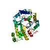
| ||||||||
|---|---|---|---|---|---|---|---|---|---|
| 1 |
| ||||||||
| Unit cell |
| ||||||||
| Atom site foot note | 1: CIS PROLINE - PRO 202 / 2: CIS PROLINE - PRO 226 3: TRP 313 - ASN 314 OMEGA = 1.33 PEPTIDE BOND DEVIATES SIGNIFICANTLY FROM TRANS CONFORMATION |
-Components
| #1: Protein | Mass: 40947.352 Da / Num. of mol.: 1 / Mutation: E140Q Source method: isolated from a genetically manipulated source Source: (gene. exp.) Clostridium thermocellum (bacteria) / Strain: NCIB 10682 / Plasmid: PCT306 (PUC9) / Production host: References: UniProt: P07985, UniProt: P0C2S3*PLUS, cellulase |
|---|---|
| #2: Water | ChemComp-HOH /  Mass: 18.015 Da / Num. of mol.: 288 / Source method: isolated from a natural source / Formula: H2O Mass: 18.015 Da / Num. of mol.: 288 / Source method: isolated from a natural source / Formula: H2O |
-Experimental details
-Experiment
| Experiment | Method: X-RAY DIFFRACTION |
|---|
- Sample preparation
| Crystal | Density Matthews: 2.31 Å3/Da / Density % sol: 46.87 % | ||||||||||||||||||||||||
|---|---|---|---|---|---|---|---|---|---|---|---|---|---|---|---|---|---|---|---|---|---|---|---|---|---|
| Crystal grow | *PLUS pH: 7.2 / Method: unknown | ||||||||||||||||||||||||
| Components of the solutions | *PLUS
|
-Data collection
| Diffraction source | Source: SYNCHROTRON / Site: EMBL/DESY, HAMBURG  / Beamline: X11 / Wavelength: 0.927 / Beamline: X11 / Wavelength: 0.927 |
|---|---|
| Detector | Type: MARRESEARCH / Detector: IMAGE PLATE / Date: Jun 1, 1995 |
| Radiation | Monochromatic (M) / Laue (L): M / Scattering type: x-ray |
| Radiation wavelength | Wavelength: 0.927 Å / Relative weight: 1 |
| Reflection | Resolution: 1.8→50 Å / Num. obs: 34277 / % possible obs: 97.8 % / Redundancy: 5.7 % / Rmerge(I) obs: 0.086 |
| Reflection | *PLUS Num. measured all: 195009 / Rmerge(I) obs: 0.086 |
- Processing
| Software |
| ||||||||||||||||||||||||||||||||||||||||||||||||||||||||||||
|---|---|---|---|---|---|---|---|---|---|---|---|---|---|---|---|---|---|---|---|---|---|---|---|---|---|---|---|---|---|---|---|---|---|---|---|---|---|---|---|---|---|---|---|---|---|---|---|---|---|---|---|---|---|---|---|---|---|---|---|---|---|
| Refinement | Resolution: 1.9→10 Å
| ||||||||||||||||||||||||||||||||||||||||||||||||||||||||||||
| Displacement parameters | Biso mean: 25.1 Å2 | ||||||||||||||||||||||||||||||||||||||||||||||||||||||||||||
| Refinement step | Cycle: LAST / Resolution: 1.9→10 Å
| ||||||||||||||||||||||||||||||||||||||||||||||||||||||||||||
| Refine LS restraints |
| ||||||||||||||||||||||||||||||||||||||||||||||||||||||||||||
| Software | *PLUS Name: X-PLOR / Classification: refinement | ||||||||||||||||||||||||||||||||||||||||||||||||||||||||||||
| Refinement | *PLUS | ||||||||||||||||||||||||||||||||||||||||||||||||||||||||||||
| Solvent computation | *PLUS | ||||||||||||||||||||||||||||||||||||||||||||||||||||||||||||
| Displacement parameters | *PLUS | ||||||||||||||||||||||||||||||||||||||||||||||||||||||||||||
| Refine LS restraints | *PLUS
|
