- PDB-1cap: CONFORMATION AND MOLECULAR ORGANIZATION IN FIBERS OF THE CAPSULAR... -
+
Open data
ID or keywords:
Loading...
-
Basic information
Entry
Database: PDB / ID: 1cap
Title
CONFORMATION AND MOLECULAR ORGANIZATION IN FIBERS OF THE CAPSULAR POLYSACCHARIDE FROM ESCHERICHIA COLI M41 MUTANT
Components
(0) x alpha-D-mannopyranose-(1-3)-beta-D-glucopyranose-(1-3)-[4,6-O-[(1S)-1-carboxyethylidene]-beta-D-glucopyranose-(1-2)-alpha-D-mannopyranose-(1-4)]beta-D-glucopyranuronic acid-(1-3)-beta-D-galactopyranose-(1-2)-alpha-D-mannopyranose-(1-3)-beta-D-glucopyranose-(1-3)-[4,6-O-[(1S)-1-carboxyethylidene]-beta-D-glucopyranose-(1-2)-alpha-D-mannopyranose-(1-4)]beta-D-glucopyranuronic acid-(1-3)-beta-D-galactopyranose
A 21 SCREW AXIS PARALLEL TO Z PASSES THROUGH X=0,Y=0 AND THE 21 AXIS PARALLEL TO X IS IN THE Z=0 PLANE. THE SCREW AXIS PARALLEL TO Z WAS USED TO GENERATE THE TWELVE-RESIDUE SEGMENT FROM THE PUBLISHED COORDINATES FOR THE SIX-RESIDUE ASYMMETRIC UNIT.
Highest resolution: 3 Å Details: THE SPACE GROUP USED IN THE ANALYSIS OF THIS STRUCTURE IS P 21 21 21 IN A NON-STANDARD SETTING. THE EQUIPOINTS FOR THIS GROUP IN THE SETTING USED ARE- X,Y,Z -X,-Y,1/2+Z -X,1/2+Y,1/2-Z 1/2+X,1/2-Y,-Z
Refinement step
Cycle: LAST / Highest resolution: 3 Å
Protein
Nucleic acid
Ligand
Solvent
Total
Num. atoms
0
0
144
0
144
+
About Yorodumi
-
News
-
Feb 9, 2022. New format data for meta-information of EMDB entries
New format data for meta-information of EMDB entries
Version 3 of the EMDB header file is now the official format.
The previous official version 1.9 will be removed from the archive.
In the structure databanks used in Yorodumi, some data are registered as the other names, "COVID-19 virus" and "2019-nCoV". Here are the details of the virus and the list of structure data.
Jan 31, 2019. EMDB accession codes are about to change! (news from PDBe EMDB page)
EMDB accession codes are about to change! (news from PDBe EMDB page)
The allocation of 4 digits for EMDB accession codes will soon come to an end. Whilst these codes will remain in use, new EMDB accession codes will include an additional digit and will expand incrementally as the available range of codes is exhausted. The current 4-digit format prefixed with “EMD-” (i.e. EMD-XXXX) will advance to a 5-digit format (i.e. EMD-XXXXX), and so on. It is currently estimated that the 4-digit codes will be depleted around Spring 2019, at which point the 5-digit format will come into force.
The EM Navigator/Yorodumi systems omit the EMD- prefix.
Related info.:Q: What is EMD? / ID/Accession-code notation in Yorodumi/EM Navigator
Yorodumi is a browser for structure data from EMDB, PDB, SASBDB, etc.
This page is also the successor to EM Navigator detail page, and also detail information page/front-end page for Omokage search.
The word "yorodu" (or yorozu) is an old Japanese word meaning "ten thousand". "mi" (miru) is to see.
Related info.:EMDB / PDB / SASBDB / Comparison of 3 databanks / Yorodumi Search / Aug 31, 2016. New EM Navigator & Yorodumi / Yorodumi Papers / Jmol/JSmol / Function and homology information / Changes in new EM Navigator and Yorodumi
 Movie
Movie Controller

 Yorodumi
 Open data
 Basic information
 Components
 Keywords
 FIBER DIFFRACTION / Resolution: 3 Å
  Authors
 Citation
 Structure visualization
 Molmil
 Downloads & links
 Other downloads
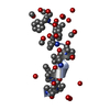
 PDBj
PDBj Assembly
 Sample preparation
 Processing