 Movie
Movie Controller

[English] 日本語
 Yorodumi
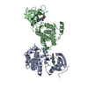
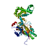
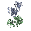
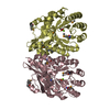
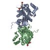
- PDB-2zd7: The structure of VPS75 (Vacuolar protein sorting-associated prote... -
+ Open data

- Basic information
| Entry | Database: PDB / ID: 2zd7 | ||||||
|---|---|---|---|---|---|---|---|
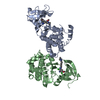
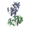
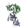
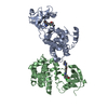
| Title | The structure of VPS75 (Vacuolar protein sorting-associated protein 75) | ||||||
 Components Components |
| ||||||
 Keywords Keywords | PROTEIN TRANSPORT / Histone chaperone / vps75 / NAP1 / Nucleus / Phosphoprotein / Transport | ||||||
| Function / homology |  Function and homology information Function and homology informationH3 histone acetyltransferase complex / acetyltransferase activator activity / DNA replication-dependent chromatin assembly / double-strand break repair via nonhomologous end joining / protein transport / nucleosome assembly / chromatin organization / histone binding / chromatin binding / chromatin ...H3 histone acetyltransferase complex / acetyltransferase activator activity / DNA replication-dependent chromatin assembly / double-strand break repair via nonhomologous end joining / protein transport / nucleosome assembly / chromatin organization / histone binding / chromatin binding / chromatin / identical protein binding / nucleus / cytosol Similarity search - Function | ||||||
| Biological species |  synthetic construct (others) | ||||||
| Method |  X-RAY DIFFRACTION / SYNCHROTRON / MAD / Resolution: 1.85 Å X-RAY DIFFRACTION / SYNCHROTRON / MAD / Resolution: 1.85 Å | ||||||
 Authors Authors | Park, Y.J. / Luger, K. | ||||||
 Citation Citation | Journal: Nat.Struct.Mol.Biol. / Year: 2008 Title: Histone chaperone specificity in Rtt109 activation. Authors: Park, Y.J. / Sudhoff, K.B. / Andrews, A.J. / Stargell, L.A. / Luger, K. #1: Journal: NAT.STRUCT.MOL.BIOL. / Year: 2008Title: Histone chaperone specificity in Rtt109 activation Authors: Park, Y.J. / Sudhoff, K.B. / Andrews, A.J. / Stargell, L.A. / Luger, K. | ||||||
| History |
|
- Structure visualization
| Structure viewer | Molecule: MolmilJmol/JSmol |
|---|
- Downloads & links
-Download
| PDBx/mmCIF format | 2zd7.cif.gz | 108.2 KB | Display | PDBx/mmCIF format |
|---|---|---|---|---|
| PDB format | pdb2zd7.ent.gz | 84.3 KB | Display | PDB format |
| PDBx/mmJSON format | 2zd7.json.gz | Tree view | PDBx/mmJSON format | |
| Others |  Other downloads Other downloads |
-Validation report
| Arichive directory | https://data.pdbj.org/pub/pdb/validation_reports/zd/2zd7ftp://data.pdbj.org/pub/pdb/validation_reports/zd/2zd7 | HTTPS FTP |
|---|
-Related structure data
| Similar structure data |
|---|

-Links
 PDBj
PDBj- Assembly
| Deposited unit | 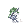
| ||||||||
|---|---|---|---|---|---|---|---|---|---|
| 1 | 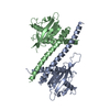
| ||||||||
| 2 | 
| ||||||||
| Unit cell |
|
-Components
| #1: Protein | Mass: 30656.084 Da / Num. of mol.: 2 Source method: isolated from a genetically manipulated source Source: (gene. exp.) Gene: VPS75 / Plasmid: PET28A / Production host:  #2: Protein/peptide | | Mass: 1416.442 Da / Num. of mol.: 1 / Source method: obtained synthetically / Source: (synth.) synthetic construct (others) #3: Water | ChemComp-HOH / |  Mass: 18.015 Da / Num. of mol.: 266 / Source method: isolated from a natural source / Formula: H2O Mass: 18.015 Da / Num. of mol.: 266 / Source method: isolated from a natural source / Formula: H2O |
|---|
-Experimental details
-Experiment
| Experiment | Method: X-RAY DIFFRACTION / Number of used crystals: 1 |
|---|
- Sample preparation
| Crystal | Density Matthews: 2.35 Å3/Da / Density % sol: 47.55 % |
|---|---|
| Crystal grow | Temperature: 292 K / Method: vapor diffusion, sitting drop / pH: 7.5 Details: PEG4000, pH 7.5, VAPOR DIFFUSION, SITTING DROP, temperature 292K |
-Data collection
| Diffraction | Mean temperature: 298 K |
|---|---|
| Diffraction source | Source: SYNCHROTRON / Site: ALS  / Beamline: 4.2.2 / Wavelength: 1.1 Å / Beamline: 4.2.2 / Wavelength: 1.1 Å |
| Detector | Detector: CCD |
| Radiation | Protocol: MAD / Monochromatic (M) / Laue (L): M / Scattering type: x-ray |
| Radiation wavelength | Wavelength: 1.1 Å / Relative weight: 1 |
| Reflection | Resolution: 1.85→50 Å / Num. all: 49841 / Num. obs: 48480 / % possible obs: 95.3 % / Redundancy: 8 % / Rmerge(I) obs: 0.081 / Net I/σ(I): 11.4 |
| Reflection shell | Resolution: 1.85→50 Å / Redundancy: 7.6 % / Rmerge(I) obs: 0.421 / Mean I/σ(I) obs: 4.1 / % possible all: 93 |
- Processing
| Software |
| ||||||||||||||||||||
|---|---|---|---|---|---|---|---|---|---|---|---|---|---|---|---|---|---|---|---|---|---|
| Refinement | Method to determine structure: MAD / Resolution: 1.85→50 Å / σ(F): 0 / σ(I): 0
| ||||||||||||||||||||
| Refinement step | Cycle: LAST / Resolution: 1.85→50 Å
| ||||||||||||||||||||
| Refine LS restraints |
|
