 Movie
Movie Controller

[English] 日本語
 Yorodumi
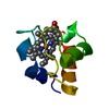
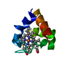
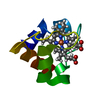
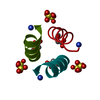
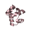
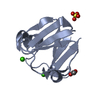
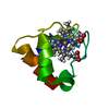
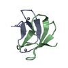
- PDB-1c75: 0.97 A "AB INITIO" CRYSTAL STRUCTURE OF CYTOCHROME C-553 FROM BAC... -
+ Open data

- Basic information
| Entry | Database: PDB / ID: 1c75 | ||||||||||||
|---|---|---|---|---|---|---|---|---|---|---|---|---|---|
| Title | 0.97 A "AB INITIO" CRYSTAL STRUCTURE OF CYTOCHROME C-553 FROM BACILLUS PASTEURII | ||||||||||||
 Components Components | CYTOCHROME C-553 | ||||||||||||
 Keywords Keywords | ELECTRON TRANSPORT / C-553 / HEME / CYTOCHROME / BACILLUS PASTEURII / AB INITIO / ATOMIC RESOLUTION | ||||||||||||
| Function / homology |  Function and homology information Function and homology informationelectron transfer activity / iron ion binding / heme binding / plasma membrane Similarity search - Function | ||||||||||||
| Biological species |  Sporosarcina pasteurii (bacteria) Sporosarcina pasteurii (bacteria) | ||||||||||||
| Method |  X-RAY DIFFRACTION / SYNCHROTRON / OTHER / Resolution: 0.97 Å X-RAY DIFFRACTION / SYNCHROTRON / OTHER / Resolution: 0.97 Å | ||||||||||||
 Authors Authors | Benini, S. / Ciurli, S. / Rypniewski, W.R. / Wilson, K.S. | ||||||||||||
 Citation Citation | Journal: Biochemistry / Year: 2000 Title: Crystal structure of oxidized Bacillus pasteurii cytochrome c553 at 0.97-A resolution. Authors: Benini, S. / Gonzalez, A. / Rypniewski, W.R. / Wilson, K.S. / Van Beeumen, J.J. / Ciurli, S. #1: Journal: Biochem.Biophys.Res.Commun. / Year: 1999Title: Cytochrome C-553 from the Alkalophilic Bacterium Bacillus Pasteurii Has the Primary Structure Characteristics of a Lipoprotein Authors: Vandenberghe, I.H.M. / Guisez, Y. / Ciurli, S. / Benini, S. / Van-Beeumen, J.J. #2: Journal: J.Biol.Inorg.Chem. / Year: 1998Title: Modulation of Bacillus Pasteurii Cytochrome C553 Reduction Potential by Structural and Solution Parameters Authors: Benini, S. / Borsari, M. / Ciurli, S. / Dikiy, A. / Lamborghini, M. #3: Journal: Proteins / Year: 1997Title: Crystals of Cytochrome C-553 from Bacillus Pasteurii Show Diffraction to 0.97 A Resolution Authors: Benini, S. / Ciurli, S. / Rypniewski, W.R. / Wilson, K.S. | ||||||||||||
| History |
|
- Structure visualization
| Structure viewer | Molecule: MolmilJmol/JSmol |
|---|
- Downloads & links
-Download
| PDBx/mmCIF format | 1c75.cif.gz | 64.9 KB | Display | PDBx/mmCIF format |
|---|---|---|---|---|
| PDB format | pdb1c75.ent.gz | 46.9 KB | Display | PDB format |
| PDBx/mmJSON format | 1c75.json.gz | Tree view | PDBx/mmJSON format | |
| Others |  Other downloads Other downloads |
-Validation report
| Summary document | 1c75_validation.pdf.gz | 772.7 KB | Display | wwPDB validaton report |
|---|---|---|---|---|
| Full document | 1c75_full_validation.pdf.gz | 774.7 KB | Display | |
| Data in XML | 1c75_validation.xml.gz | 7.3 KB | Display | |
| Data in CIF | 1c75_validation.cif.gz | 9.5 KB | Display | |
| Arichive directory | https://data.pdbj.org/pub/pdb/validation_reports/c7/1c75ftp://data.pdbj.org/pub/pdb/validation_reports/c7/1c75 | HTTPS FTP |
-Related structure data

-Links
 PDBj
PDBj


- Assembly
| Deposited unit | 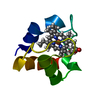
| ||||||||
|---|---|---|---|---|---|---|---|---|---|
| 1 |
| ||||||||
| Unit cell |
|
-Components
| #1: Protein | Mass: 7115.937 Da / Num. of mol.: 1 / Source method: isolated from a natural source / Details: DEUTSCHE SAMMLUNG VON MIKROORGANISMEN (DSM) / Source: (natural) Sporosarcina pasteurii (bacteria) / Cellular location: CYTOPLASM / Strain: DSM 33 / References: UniProt: P82599 |
|---|---|
| #2: Chemical | ChemComp-HEC /   Mass: 618.503 Da / Num. of mol.: 1 / Source method: obtained synthetically / Formula: C34H34FeN4O4 Mass: 618.503 Da / Num. of mol.: 1 / Source method: obtained synthetically / Formula: C34H34FeN4O4 |
| #3: Water | ChemComp-HOH /  Mass: 18.015 Da / Num. of mol.: 125 / Source method: isolated from a natural source / Formula: H2O Mass: 18.015 Da / Num. of mol.: 125 / Source method: isolated from a natural source / Formula: H2O |
| Has protein modification | Y |
-Experimental details
-Experiment
| Experiment | Method: X-RAY DIFFRACTION / Number of used crystals: 1 |
|---|
- Sample preparation
| Crystal | Density Matthews: 2.08 Å3/Da / Density % sol: 38 % / Description: THE STRUCTURE WAS SOLVED "AB INITIO" | |||||||||||||||||||||||||
|---|---|---|---|---|---|---|---|---|---|---|---|---|---|---|---|---|---|---|---|---|---|---|---|---|---|---|
| Crystal grow | pH: 5 Details: 3.2 M AMMONIUM SULPHATE IN 100 MM SODIUM CITRATE BUFFER PH 5.0 | |||||||||||||||||||||||||
| Crystal | *PLUS Density % sol: 41 % | |||||||||||||||||||||||||
| Crystal grow | *PLUS Temperature: 20 ℃ / pH: 8 / Method: vapor diffusion, hanging drop | |||||||||||||||||||||||||
| Components of the solutions | *PLUS
|
-Data collection
| Diffraction | Mean temperature: 100 K |
|---|---|
| Diffraction source | Source: SYNCHROTRON / Site: EMBL/DESY, HAMBURG  / Beamline: BW7B / Wavelength: 0.885 / Beamline: BW7B / Wavelength: 0.885 |
| Detector | Type: MARRESEARCH / Detector: IMAGE PLATE / Date: May 23, 1996 / Details: SEGMENTED MIRROR |
| Radiation | Monochromator: SI(111) / Protocol: SINGLE WAVELENGTH / Monochromatic (M) / Laue (L): M / Scattering type: x-ray |
| Radiation wavelength | Wavelength: 0.885 Å / Relative weight: 1 |
| Reflection | Resolution: 0.97→20 Å / Num. obs: 38959 / % possible obs: 99.9 % / Observed criterion σ(I): -3 / Redundancy: 5.46 % / Rmerge(I) obs: 0.074 / Rsym value: 0.074 / Net I/σ(I): 17.16 |
| Reflection shell | Resolution: 0.97→0.99 Å / Redundancy: 2.72 % / Rmerge(I) obs: 0.242 / Mean I/σ(I) obs: 4.18 / Rsym value: 0.242 / % possible all: 99.6 |
| Reflection | *PLUS Lowest resolution: 20 Å / Redundancy: 5.47 % / Num. measured all: 212694 / Rmerge(I) obs: 0.074 |
| Reflection shell | *PLUS |
- Processing
| Software |
| |||||||||||||||||||||||||||||||||
|---|---|---|---|---|---|---|---|---|---|---|---|---|---|---|---|---|---|---|---|---|---|---|---|---|---|---|---|---|---|---|---|---|---|---|
| Refinement | Method to determine structure: OTHER / Resolution: 0.97→20 Å / Num. parameters: 6286 / Num. restraintsaints: 7877 / σ(F): 0 / Stereochemistry target values: ENGH AND HUBER
| |||||||||||||||||||||||||||||||||
| Solvent computation | Solvent model: MOEWS & KRETSINGER, J.MOL.BIOL.91(1973)201 | |||||||||||||||||||||||||||||||||
| Refine analyze | Num. disordered residues: 14 / Occupancy sum hydrogen: 486 / Occupancy sum non hydrogen: 661.12 | |||||||||||||||||||||||||||||||||
| Refinement step | Cycle: LAST / Resolution: 0.97→20 Å
| |||||||||||||||||||||||||||||||||
| Refine LS restraints |
| |||||||||||||||||||||||||||||||||
| Software | *PLUS Name: SHELXL-97 / Classification: refinement | |||||||||||||||||||||||||||||||||
| Refinement | *PLUS Lowest resolution: 20 Å / σ(F): 0 / % reflection Rfree: 5 % | |||||||||||||||||||||||||||||||||
| Solvent computation | *PLUS | |||||||||||||||||||||||||||||||||
| Displacement parameters | *PLUS Biso mean: 14.7 Å2 | |||||||||||||||||||||||||||||||||
| Refine LS restraints | *PLUS
|
