 Movie
Movie Controller


+ Open data

- Basic information
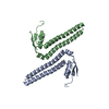
| Entry | Database: PDB / ID: 1c3g | ||||||
|---|---|---|---|---|---|---|---|
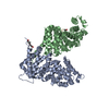
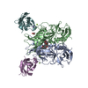
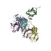
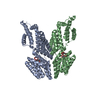
| Title | S. CEREVISIAE HEAT SHOCK PROTEIN 40 SIS1 | ||||||
 Components Components | HEAT SHOCK PROTEIN 40 | ||||||
 Keywords Keywords | CHAPERONE / BETA SHEETS / SHORT HELICES | ||||||
| Function / homology |  Function and homology information Function and homology informationdetection of misfolded protein / tRNA import into nucleus / misfolded protein transport / mitochondria-associated ubiquitin-dependent protein catabolic process / nuclear protein quality control by the ubiquitin-proteasome system / misfolded protein binding / cellular response to starvation / translational initiation / : / cellular response to heat ...detection of misfolded protein / tRNA import into nucleus / misfolded protein transport / mitochondria-associated ubiquitin-dependent protein catabolic process / nuclear protein quality control by the ubiquitin-proteasome system / misfolded protein binding / cellular response to starvation / translational initiation / : / cellular response to heat / protein-folding chaperone binding / protein folding / cytosolic small ribosomal subunit / nucleolus / DNA binding / nucleus / cytosol Similarity search - Function | ||||||
| Biological species |  | ||||||
| Method |  X-RAY DIFFRACTION / SYNCHROTRON / Resolution: 2.7 Å X-RAY DIFFRACTION / SYNCHROTRON / Resolution: 2.7 Å | ||||||
 Authors Authors | Sha, B. / Lee, S. / Cyr, D. | ||||||
 Citation Citation | Journal: Structure / Year: 2000 Title: The crystal structure of the peptide-binding fragment from the yeast Hsp40 protein Sis1. Authors: Sha, B. / Lee, S. / Cyr, D.M. | ||||||
| History |
|
- Structure visualization
| Structure viewer | Molecule: MolmilJmol/JSmol |
|---|
- Downloads & links
-Download
| PDBx/mmCIF format | 1c3g.cif.gz | 45.5 KB | Display | PDBx/mmCIF format |
|---|---|---|---|---|
| PDB format | pdb1c3g.ent.gz | 32.9 KB | Display | PDB format |
| PDBx/mmJSON format | 1c3g.json.gz | Tree view | PDBx/mmJSON format | |
| Others |  Other downloads Other downloads |
-Validation report
| Arichive directory | https://data.pdbj.org/pub/pdb/validation_reports/c3/1c3gftp://data.pdbj.org/pub/pdb/validation_reports/c3/1c3g | HTTPS FTP |
|---|
-Related structure data
| Similar structure data |
|---|


-Links
 PDBj
PDBj


- Assembly
| Deposited unit | 
| ||||||||
|---|---|---|---|---|---|---|---|---|---|
| 1 | 
| ||||||||
| Unit cell |
| ||||||||
| Details | the biological assembly is a homodimer constructed by a crystallographic 2-fold. |
-Components
| #1: Protein | Mass: 19097.801 Da / Num. of mol.: 1 / Fragment: SIS1 C-TERMINAL PEPTIDE-BINDING DOMAIN Source method: isolated from a genetically manipulated source Details: MOLECULAR CHAPERONE Source: (gene. exp.) Plasmid: PET9D / Production host:  |
|---|---|
| #2: Water | ChemComp-HOH /  Mass: 18.015 Da / Num. of mol.: 33 / Source method: isolated from a natural source / Formula: H2O Mass: 18.015 Da / Num. of mol.: 33 / Source method: isolated from a natural source / Formula: H2O |
-Experimental details
-Experiment
| Experiment | Method: X-RAY DIFFRACTION / Number of used crystals: 1 |
|---|
- Sample preparation
| Crystal | Density Matthews: 2.82 Å3/Da / Density % sol: 56.44 % | ||||||||||||||||||||||||||||||||||||||||||
|---|---|---|---|---|---|---|---|---|---|---|---|---|---|---|---|---|---|---|---|---|---|---|---|---|---|---|---|---|---|---|---|---|---|---|---|---|---|---|---|---|---|---|---|
| Crystal grow | Temperature: 298 K / Method: vapor diffusion, hanging drop / pH: 7.2 Details: PEG 3350 20%, pH 7.2, VAPOR DIFFUSION, HANGING DROP, temperature 25K | ||||||||||||||||||||||||||||||||||||||||||
| Crystal grow | *PLUS Details: Sha, B.D., (1999) Acta Crystallogr, D55, 1234. | ||||||||||||||||||||||||||||||||||||||||||
| Components of the solutions | *PLUS
|
-Data collection
| Diffraction | Mean temperature: 100 K |
|---|---|
| Diffraction source | Source: SYNCHROTRON / Site: SSRL  / Beamline: BL7-1 / Wavelength: 1.08 / Beamline: BL7-1 / Wavelength: 1.08 |
| Detector | Type: MARRESEARCH / Detector: IMAGE PLATE / Date: Jan 1, 1998 |
| Radiation | Protocol: SINGLE WAVELENGTH / Monochromatic (M) / Laue (L): M / Scattering type: x-ray |
| Radiation wavelength | Wavelength: 1.08 Å / Relative weight: 1 |
| Reflection | Resolution: 2.7→30 Å / Num. all: 6400 / Num. obs: 6347 / % possible obs: 97 % / Observed criterion σ(F): 2 / Observed criterion σ(I): 2 / Redundancy: 5.5 % / Biso Wilson estimate: 70 Å2 / Rmerge(I) obs: 0.045 / Net I/σ(I): 20.5 |
| Reflection shell | Resolution: 2.7→2.9 Å / Redundancy: 3 % / Rmerge(I) obs: 0.35 / Num. unique all: 6347 / % possible all: 98 |
| Reflection | *PLUS % possible obs: 97 % / Rmerge(I) obs: 0.045 |
| Reflection shell | *PLUS % possible obs: 99.5 % / Rmerge(I) obs: 0.377 / Mean I/σ(I) obs: 6.17 |
- Processing
| Software |
| |||||||||||||||||||||||||
|---|---|---|---|---|---|---|---|---|---|---|---|---|---|---|---|---|---|---|---|---|---|---|---|---|---|---|
| Refinement | Resolution: 2.7→30 Å / σ(F): 0 / σ(I): 0 / Stereochemistry target values: Engh & Huber / Details: Simulated annealing
| |||||||||||||||||||||||||
| Refinement step | Cycle: LAST / Resolution: 2.7→30 Å
| |||||||||||||||||||||||||
| Refine LS restraints |
| |||||||||||||||||||||||||
| Software | *PLUS Name: CNS / Classification: refinement | |||||||||||||||||||||||||
| Refinement | *PLUS Lowest resolution: 30 Å / Num. reflection Rfree: 508 / Rfactor obs: 0.265 / Rfactor Rfree: 0.311 / Rfactor Rwork: 0.265 | |||||||||||||||||||||||||
| Solvent computation | *PLUS | |||||||||||||||||||||||||
| Displacement parameters | *PLUS | |||||||||||||||||||||||||
| Refine LS restraints | *PLUS
| |||||||||||||||||||||||||
| LS refinement shell | *PLUS Rfactor Rfree: 0.42 / Rfactor Rwork: 0.382 |
