 Movie
Movie Controller

[English] 日本語
 Yorodumi
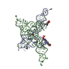
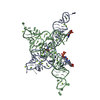
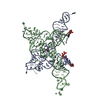
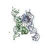
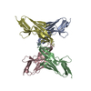
- PDB-5uh7: Crystal structure of beta'MtbSI of Mycobacterium tuberculosis RNA... -
+ Open data

- Basic information
| Entry | Database: PDB / ID: 5uh7 | ||||||
|---|---|---|---|---|---|---|---|
| Title | Crystal structure of beta'MtbSI of Mycobacterium tuberculosis RNA polymerase | ||||||
 Components Components | DNA-directed RNA polymerase subunit beta' | ||||||
 Keywords Keywords | TRANSCRIPTION / RNA polymerase complex / DNA / RNA | ||||||
| Function / homology |  Function and homology information Function and homology informationAntimicrobial action and antimicrobial resistance in Mtb / peptidoglycan-based cell wall / DNA-directed RNA polymerase complex / DNA-directed RNA polymerase / DNA-directed RNA polymerase activity / DNA-templated transcription / magnesium ion binding / DNA binding / zinc ion binding / plasma membrane / cytosol Similarity search - Function | ||||||
| Biological species |   Mycobacterium tuberculosis (bacteria) Mycobacterium tuberculosis (bacteria) | ||||||
| Method |  X-RAY DIFFRACTION / SYNCHROTRON / Resolution: 2.199 Å X-RAY DIFFRACTION / SYNCHROTRON / Resolution: 2.199 Å | ||||||
 Authors Authors | Lin, W. / Das, K. / Feng, Y. / Ebright, R.H. | ||||||
 Citation Citation | Journal: Mol. Cell / Year: 2017 Title: Structural Basis of Mycobacterium tuberculosis Transcription and Transcription Inhibition. Authors: Lin, W. / Mandal, S. / Degen, D. / Liu, Y. / Ebright, Y.W. / Li, S. / Feng, Y. / Zhang, Y. / Mandal, S. / Jiang, Y. / Liu, S. / Gigliotti, M. / Talaue, M. / Connell, N. / Das, K. / Arnold, E. / Ebright, R.H. | ||||||
| History |
|
- Structure visualization
| Structure viewer | Molecule: MolmilJmol/JSmol |
|---|
- Downloads & links
-Download
| PDBx/mmCIF format | 5uh7.cif.gz | 120.7 KB | Display | PDBx/mmCIF format |
|---|---|---|---|---|
| PDB format | pdb5uh7.ent.gz | 94.6 KB | Display | PDB format |
| PDBx/mmJSON format | 5uh7.json.gz | Tree view | PDBx/mmJSON format | |
| Others |  Other downloads Other downloads |
-Validation report
| Arichive directory | https://data.pdbj.org/pub/pdb/validation_reports/uh/5uh7ftp://data.pdbj.org/pub/pdb/validation_reports/uh/5uh7 | HTTPS FTP |
|---|
-Related structure data
| Related structure data |  5uh5C  5uh6C  5uh8C  5uh9C  5uhaC  5uhbC  5uhcC  5uhdC  5uheC  5uhfC  5uhgC C: citing same article ( |
|---|---|
| Similar structure data |


-Links
 PDBj
PDBj


- Assembly
| Deposited unit | 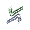
| ||||||||
|---|---|---|---|---|---|---|---|---|---|
| 1 |
| ||||||||
| Unit cell |
|
-Components
| #1: Protein | Mass: 19512.736 Da / Num. of mol.: 2 Source method: isolated from a genetically manipulated source Source: (gene. exp.) Mycobacterium tuberculosis (strain ATCC 25618 / H37Rv) (bacteria)Strain: ATCC 25618 / H37Rv / Gene: rpoC, Rv0668, MTCI376.07c Production host: References: UniProt: P9WGY7, DNA-directed RNA polymerase #2: Water | ChemComp-HOH / |  Mass: 18.015 Da / Num. of mol.: 43 / Source method: isolated from a natural source / Formula: H2O Mass: 18.015 Da / Num. of mol.: 43 / Source method: isolated from a natural source / Formula: H2O |
|---|
-Experimental details
-Experiment
| Experiment | Method: X-RAY DIFFRACTION / Number of used crystals: 1 |
|---|
- Sample preparation
| Crystal | Density Matthews: 2.73 Å3/Da / Density % sol: 54.97 % |
|---|---|
| Crystal grow | Temperature: 295 K / Method: vapor diffusion, hanging drop / pH: 5.5 Details: 100 mM sodium citrate tribasic dihydrate, pH 5.5, and 22% PEG1000 |
-Data collection
| Diffraction | Mean temperature: 100 K | |||||||||||||||||||||||||||||||||||||||||||||||||||||||||||||||||||||||||||||||||||||||||||||||||||
|---|---|---|---|---|---|---|---|---|---|---|---|---|---|---|---|---|---|---|---|---|---|---|---|---|---|---|---|---|---|---|---|---|---|---|---|---|---|---|---|---|---|---|---|---|---|---|---|---|---|---|---|---|---|---|---|---|---|---|---|---|---|---|---|---|---|---|---|---|---|---|---|---|---|---|---|---|---|---|---|---|---|---|---|---|---|---|---|---|---|---|---|---|---|---|---|---|---|---|---|---|
| Diffraction source | Source: SYNCHROTRON / Site: APS  / Beamline: 19-ID / Wavelength: 0.97925 Å / Beamline: 19-ID / Wavelength: 0.97925 Å | |||||||||||||||||||||||||||||||||||||||||||||||||||||||||||||||||||||||||||||||||||||||||||||||||||
| Detector | Type: DECTRIS PILATUS3 6M / Detector: PIXEL / Date: Nov 22, 2015 | |||||||||||||||||||||||||||||||||||||||||||||||||||||||||||||||||||||||||||||||||||||||||||||||||||
| Radiation | Protocol: SINGLE WAVELENGTH / Monochromatic (M) / Laue (L): M / Scattering type: x-ray | |||||||||||||||||||||||||||||||||||||||||||||||||||||||||||||||||||||||||||||||||||||||||||||||||||
| Radiation wavelength | Wavelength: 0.97925 Å / Relative weight: 1 | |||||||||||||||||||||||||||||||||||||||||||||||||||||||||||||||||||||||||||||||||||||||||||||||||||
| Reflection | Resolution: 2.2→50 Å / Num. obs: 21229 / % possible obs: 99.4 % / Redundancy: 4.3 % / Rmerge(I) obs: 0.075 / Rpim(I) all: 0.04 / Rrim(I) all: 0.086 / Χ2: 0.841 / Net I/σ(I): 11 / Num. measured all: 91896 | |||||||||||||||||||||||||||||||||||||||||||||||||||||||||||||||||||||||||||||||||||||||||||||||||||
| Reflection shell | Diffraction-ID: 1
|
- Processing
| Software |
| ||||||||||||||||||||||||
|---|---|---|---|---|---|---|---|---|---|---|---|---|---|---|---|---|---|---|---|---|---|---|---|---|---|
| Refinement | Resolution: 2.199→36.08 Å / SU ML: 0.27 / Cross valid method: FREE R-VALUE / σ(F): 1.36 / Phase error: 28.11 / Stereochemistry target values: ML
| ||||||||||||||||||||||||
| Solvent computation | Shrinkage radii: 0.9 Å / VDW probe radii: 1.11 Å / Solvent model: FLAT BULK SOLVENT MODEL | ||||||||||||||||||||||||
| Displacement parameters | Biso max: 133.98 Å2 / Biso mean: 49.2648 Å2 / Biso min: 19.16 Å2 | ||||||||||||||||||||||||
| Refinement step | Cycle: final / Resolution: 2.199→36.08 Å
|
