[English] 日本語
 Yorodumi
- PDB-1c04: IDENTIFICATION OF KNOWN PROTEIN AND RNA STRUCTURES IN A 5 A MAP O... -
+ Open data
- Basic information
| Entry | Database: PDB / ID: 1c04 | ||||||
|---|---|---|---|---|---|---|---|
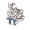
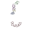
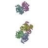
| Title | IDENTIFICATION OF KNOWN PROTEIN AND RNA STRUCTURES IN A 5 A MAP OF THE LARGE RIBOSOMAL SUBUNIT FROM HALOARCULA MARISMORTUI | ||||||
 Components Components |
| ||||||
 Keywords Keywords | RIBOSOME / LOW RESOLUTION MODEL / LARGE RIBOSOME UNIT | ||||||
| Function / homology |  Function and homology information Function and homology informationlarge ribosomal subunit / transferase activity / large ribosomal subunit rRNA binding / cytosolic large ribosomal subunit / cytoplasmic translation / rRNA binding / structural constituent of ribosome / translation / ribonucleoprotein complex Similarity search - Function | ||||||
| Biological species |  Haloarcula marismortui (Halophile) Haloarcula marismortui (Halophile) | ||||||
| Method |  X-RAY DIFFRACTION / X-RAY DIFFRACTION /  SYNCHROTRON / Resolution: 5 Å SYNCHROTRON / Resolution: 5 Å | ||||||
 Authors Authors | Ban, N. / Nissen, P. / Capel, M. / Moore, P.B. / Steitz, T.A. | ||||||
 Citation Citation |  Journal: Nature / Year: 1999 Journal: Nature / Year: 1999Title: Placement of protein and RNA structures into a 5 A-resolution map of the 50S ribosomal subunit. Authors: Ban, N. / Nissen, P. / Hansen, J. / Capel, M. / Moore, P.B. / Steitz, T.A. #1:  Journal: Cell(Cambridge,Mass.) / Year: 1998 Journal: Cell(Cambridge,Mass.) / Year: 1998Title: A 9 A resolution X-ray crystallographic map of the large ribosomal subunit Authors: Ban, N. / Freeborn, B. / Nissen, P. / Penczec, P. / Grassucci, R.A. / Sweet, R. / Frank, J. / Moore, P.B. / Steitz, T.A. | ||||||
| History |
|
- Structure visualization
| Structure viewer | Molecule:  Molmil Molmil Jmol/JSmol Jmol/JSmol |
|---|
- Downloads & links
- Download
| PDBx/mmCIF format |  1c04.cif.gz 1c04.cif.gz | 153.2 KB | Display |  PDBx/mmCIF format PDBx/mmCIF format |
|---|---|---|---|---|
| PDB format |  pdb1c04.ent.gz pdb1c04.ent.gz | 114.3 KB | Display |  PDB format PDB format |
| PDBx/mmJSON format |  1c04.json.gz 1c04.json.gz | Tree view |  PDBx/mmJSON format PDBx/mmJSON format | |
| Others |  Other downloads Other downloads |
-Validation report
| Arichive directory |  https://data.pdbj.org/pub/pdb/validation_reports/c0/1c04 https://data.pdbj.org/pub/pdb/validation_reports/c0/1c04 ftp://data.pdbj.org/pub/pdb/validation_reports/c0/1c04 ftp://data.pdbj.org/pub/pdb/validation_reports/c0/1c04 | HTTPS FTP |
|---|
-Related structure data
| Related structure data | |
|---|---|
| Similar structure data |
- Links
Links
- Assembly
| Deposited unit | 
| ||||||||||
|---|---|---|---|---|---|---|---|---|---|---|---|
| 1 |
| ||||||||||
| Unit cell |
|
- Components
-RNA chain , 2 types, 2 molecules EF
| #1: RNA chain | Mass: 18725.191 Da / Num. of mol.: 1 / Fragment: 23S RRNA 1151-1208 REGION / Source method: isolated from a natural source / Details: RNA E. COLI SEQUENCE AND MODEL / Source: (natural)  Haloarcula marismortui (Halophile) Haloarcula marismortui (Halophile) |
|---|---|
| #2: RNA chain | Mass: 9376.676 Da / Num. of mol.: 1 / Fragment: 23S RRNA HELIX 95 / Source method: isolated from a natural source / Details: RNA RAT SEQUENCE AND MODEL / Source: (natural)  Haloarcula marismortui (Halophile) Haloarcula marismortui (Halophile) |
-RIBOSOMAL PROTEIN ... , 4 types, 4 molecules ABCD
| #3: Protein | Mass: 14930.819 Da / Num. of mol.: 1 / Fragment: CENTRAL RNA-BINDING DOMAINS / Source method: isolated from a natural source Details: MODELED BY ANALOGOUS PROTEIN OF B. STEAROTHERMOPHILUS TAKEN FROM PDB ENTRY 1RL2 Source: (natural)  Haloarcula marismortui (Halophile) / References: UniProt: P04257 Haloarcula marismortui (Halophile) / References: UniProt: P04257 |
|---|---|
| #4: Protein | Mass: 19202.123 Da / Num. of mol.: 1 / Source method: isolated from a natural source Details: MODELED BY ANALOGOUS PROTEIN OF B. STEAROTHERMOPHILUS TAKEN FROM PDB ENTRY 1RL6 Source: (natural)  Haloarcula marismortui (Halophile) / References: UniProt: P02391 Haloarcula marismortui (Halophile) / References: UniProt: P02391 |
| #5: Protein | Mass: 7116.356 Da / Num. of mol.: 1 / Fragment: C-TERMINAL DOMAIN / Source method: isolated from a natural source Details: MODELED BY ANALOGOUS PROTEIN OF B. STEAROTHERMOPHILUS TAKEN FROM PDB ENTRY 1QA6 Source: (natural)  Haloarcula marismortui (Halophile) / References: UniProt: P56210 Haloarcula marismortui (Halophile) / References: UniProt: P56210 |
| #6: Protein | Mass: 13369.613 Da / Num. of mol.: 1 / Source method: isolated from a natural source Details: MODELED BY ANALOGOUS PROTEIN OF B. STEAROTHERMOPHILUS TAKEN FROM PDB ENTRY 1WHI Source: (natural)  Haloarcula marismortui (Halophile) / References: UniProt: P04450 Haloarcula marismortui (Halophile) / References: UniProt: P04450 |
-Details
| Has protein modification | Y |
|---|
-Experimental details
-Experiment
| Experiment | Method:  X-RAY DIFFRACTION / Number of used crystals: 1 X-RAY DIFFRACTION / Number of used crystals: 1 |
|---|
- Sample preparation
| Crystal grow | Temperature: 292 K / Method: vapor diffusion, hanging drop / pH: 5.4 Details: PEG 6000, POTASSIUM CHLORIDE, AMMONIUM CHLORIDE, MAGNESIUM CHLORIDE, ACETATE, pH 5.4, VAPOR DIFFUSION, HANGING DROP, temperature 292K | ||||||||||||||||||||||||||||||||||||||||||||||||||||||
|---|---|---|---|---|---|---|---|---|---|---|---|---|---|---|---|---|---|---|---|---|---|---|---|---|---|---|---|---|---|---|---|---|---|---|---|---|---|---|---|---|---|---|---|---|---|---|---|---|---|---|---|---|---|---|---|
| Components of the solutions |
| ||||||||||||||||||||||||||||||||||||||||||||||||||||||
| Crystal grow | *PLUS Temperature: 19 ℃ / pH: 5.6 / Method: vapor diffusion / Details: von Bohlen, K., (1991) J. Mol. Biol., 222, 11. | ||||||||||||||||||||||||||||||||||||||||||||||||||||||
| Components of the solutions | *PLUS
|
-Data collection
| Diffraction |
| |||||||||||||||
|---|---|---|---|---|---|---|---|---|---|---|---|---|---|---|---|---|
| Diffraction source |
| |||||||||||||||
| Detector |
| |||||||||||||||
| Radiation |
| |||||||||||||||
| Radiation wavelength |
| |||||||||||||||
| Reflection | Resolution: 3.75→130 Å / Num. all: 185190 / % possible obs: 98.6 % / Observed criterion σ(F): 0 / Observed criterion σ(I): -3 / Redundancy: 5.2 % / Biso Wilson estimate: 67 Å2 / Rmerge(I) obs: 0.103 / Net I/σ(I): 13 | |||||||||||||||
| Reflection shell | Resolution: 3.75→3.81 Å / Redundancy: 4 % / Rmerge(I) obs: 0.513 / % possible all: 96 | |||||||||||||||
| Reflection | *PLUS Num. obs: 185190 / Num. measured all: 957850 | |||||||||||||||
| Reflection shell | *PLUS Mean I/σ(I) obs: 2.2 |
- Processing
| Software |
| ||||||||||||
|---|---|---|---|---|---|---|---|---|---|---|---|---|---|
| Refinement | Resolution: 5→60 Å / Num. reflection all: 77078 / Num. reflection obs: 76415 / σ(F): 2 Details: COMBINED MIRAS AND SAD PHASES WERE DETERMINED FROM ONE NATIVE AND FOUR DERIVATIVE CRYSTALS. PHASES WERE REFINED BY MULTI-CRYSTAL AVERAGING USING THREE CRYSTAL FORMS AND BY DENSITY ...Details: COMBINED MIRAS AND SAD PHASES WERE DETERMINED FROM ONE NATIVE AND FOUR DERIVATIVE CRYSTALS. PHASES WERE REFINED BY MULTI-CRYSTAL AVERAGING USING THREE CRYSTAL FORMS AND BY DENSITY MODIFICATION. RIBOSOMAL PROTEINS AND RNA FRAGMENTS WERE MANUALLY FITTED TO THE MAP CALCULATED AT 60 - 5 A RESOLUTION. NO COMPUTATIONAL REFINEMENT OF THE FITTING HAS BEEN PERFORMED. THE L6 DOMAINS HAVE BEEN MOVED RELATIVE TO EACH OTHER BY APPROXIMATELY 5 DEG. TO IMPROVE THE FIT TO DENSITY. THE L11-RNA COMPLEX HAS BEEN SLIGHTLY ADJUSTED TO OPTIMIZE THE FIT TO DENSITY OF BOTH L11 AND THE 58NT RRNA FRAGMENT SEPARATELY. | ||||||||||||
| Refinement step | Cycle: LAST / Resolution: 5→60 Å
| ||||||||||||
| Refinement | *PLUS Highest resolution: 5 Å / Lowest resolution: 60 Å / σ(F): 2 | ||||||||||||
| Solvent computation | *PLUS | ||||||||||||
| Displacement parameters | *PLUS |
 Movie
Movie Controller


 PDBj
PDBj


