 Movie
Movie Controller


+ Open data

- Basic information
| Entry | Database: PDB / ID: 1byz | |||||||||
|---|---|---|---|---|---|---|---|---|---|---|
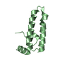
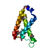
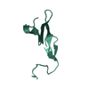
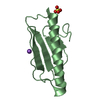
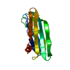
| Title | DESIGNED PEPTIDE ALPHA-1, P1 FORM | |||||||||
 Components Components | PROTEIN (SYNTHETIC DESIGNED PEPTIDE "ALPHA-1") | |||||||||
 Keywords Keywords | DE NOVO PROTEIN / HELICAL BILAYER / BIOMATERIAL | |||||||||
| Function / homology | ETHANOLAMINE Function and homology information Function and homology information | |||||||||
| Biological species | synthetic construct (others) | |||||||||
| Method |  X-RAY DIFFRACTION / SYNCHROTRON / DIRECT METHODS / Resolution: 0.9 Å X-RAY DIFFRACTION / SYNCHROTRON / DIRECT METHODS / Resolution: 0.9 Å | |||||||||
 Authors Authors | Prive, G.G. / Anderson, D.H. / Wesson, L. / Cascio, D. / Eisenberg, D. | |||||||||
 Citation Citation | Journal: Protein Sci. / Year: 1999 Title: Packed protein bilayers in the 0.90 A resolution structure of a designed alpha helical bundle. Authors: Prive, G.G. / Anderson, D.H. / Wesson, L. / Cascio, D. / Eisenberg, D. #1: Journal: Protein Sci. / Year: 1999Title: Centrosymmetric Bilayers in the 0.75A Resolution Structure of a Designed Alpha- Helical Peptide, D, L-Alpha-1 Authors: Patterson, W.R. / Anderson, D.H. / Degrado, W.F. / Cascio, D. / Eisenberg, D. #2: Journal: Science / Year: 1990Title: Crystal Structure of Alpha-1: Implications for Protein Design Authors: Hill, C.P. / Anderson, D.H. / Wesson, L. / Degrado, W.F. / Eisenberg, D. #3: Journal: Proteins / Year: 1986Title: The Design, Synthesis, and Crystallization of an Alpha-Helical Peptide Authors: Eisenberg, D. / Wilcox, W. / Eshita, S.M. / Pryciak, P.M. / Ho, S.P. / De Grado, W.F. | |||||||||
| History |
|
- Structure visualization
| Structure viewer | Molecule: MolmilJmol/JSmol |
|---|
- Downloads & links
-Download
| PDBx/mmCIF format | 1byz.cif.gz | 55.6 KB | Display | PDBx/mmCIF format |
|---|---|---|---|---|
| PDB format | pdb1byz.ent.gz | 41.6 KB | Display | PDB format |
| PDBx/mmJSON format | 1byz.json.gz | Tree view | PDBx/mmJSON format | |
| Others |  Other downloads Other downloads |
-Validation report
| Arichive directory | https://data.pdbj.org/pub/pdb/validation_reports/by/1byzftp://data.pdbj.org/pub/pdb/validation_reports/by/1byz | HTTPS FTP |
|---|
-Related structure data
| Similar structure data |
|---|


-Links
 PDBj
PDBj
- Assembly
| Deposited unit | 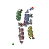
| ||||||||
|---|---|---|---|---|---|---|---|---|---|
| 1 |
| ||||||||
| Unit cell |
|
-Components
-Protein/peptide , 1 types, 4 molecules ABCD
| #1: Protein/peptide | Mass: 1441.775 Da / Num. of mol.: 4 / Source method: obtained synthetically / Details: N TERMINI ARE ACETYLATED / Source: (synth.) synthetic construct (others) |
|---|
-Non-polymers , 5 types, 37 molecules 


| #2: Chemical |  Type: L-peptide COOH carboxy terminus / Mass: 61.083 Da / Num. of mol.: 2 / Source method: obtained synthetically / Formula: C2H7NO Type: L-peptide COOH carboxy terminus / Mass: 61.083 Da / Num. of mol.: 2 / Source method: obtained synthetically / Formula: C2H7NO#3: Chemical |  Mass: 118.174 Da / Num. of mol.: 3 / Source method: obtained synthetically / Formula: C6H14O2 / Comment: precipitant*YM Mass: 118.174 Da / Num. of mol.: 3 / Source method: obtained synthetically / Formula: C6H14O2 / Comment: precipitant*YM#4: Chemical | ChemComp-CL / |  Mass: 35.453 Da / Num. of mol.: 1 / Source method: obtained synthetically / Formula: Cl Mass: 35.453 Da / Num. of mol.: 1 / Source method: obtained synthetically / Formula: Cl#5: Chemical | ChemComp-MRD / ( |  Mass: 118.174 Da / Num. of mol.: 1 / Source method: obtained synthetically / Formula: C6H14O2 / Comment: precipitant*YM Mass: 118.174 Da / Num. of mol.: 1 / Source method: obtained synthetically / Formula: C6H14O2 / Comment: precipitant*YM#6: Water | ChemComp-HOH / | Mass: 18.015 Da / Num. of mol.: 30 / Source method: isolated from a natural source / Formula: H2O |
|---|
-Details
| Has protein modification | Y |
|---|---|
| Nonpolymer details | MPD 513 IS BUILT IN TWO OVERLAPPIN |
-Experimental details
-Experiment
| Experiment | Method: X-RAY DIFFRACTION / Number of used crystals: 1 |
|---|
- Sample preparation
| Crystal | Density Matthews: 1.69 Å3/Da / Density % sol: 27.13 % / Description: SNB SUCCESS RATE WAS 5%. | |||||||||||||||||||||||||
|---|---|---|---|---|---|---|---|---|---|---|---|---|---|---|---|---|---|---|---|---|---|---|---|---|---|---|
| Crystal grow | Method: vapor diffusion, sitting drop / pH: 8 Details: 91% 2-METHYL-2,4-PENTANEDIOL, 78mM TRIETHANOLAMINE-HCL pH8, 52mM ETHANOLAMINE-HCL pH9.75, pH 8.0, VAPOR DIFFUSION, SITTING DROP | |||||||||||||||||||||||||
| Crystal grow | *PLUS Details: drop consists of equal volume of peptide and reservoir solutions | |||||||||||||||||||||||||
| Components of the solutions | *PLUS
|
-Data collection
| Diffraction | Mean temperature: 100 K |
|---|---|
| Diffraction source | Source: SYNCHROTRON / Site: NSLS  / Beamline: X12C / Wavelength: 0.9 / Beamline: X12C / Wavelength: 0.9 |
| Detector | Type: MARRESEARCH / Detector: IMAGE PLATE / Date: Nov 15, 1994 |
| Radiation | Monochromator: SI / Protocol: SINGLE WAVELENGTH / Monochromatic (M) / Laue (L): M / Scattering type: x-ray |
| Radiation wavelength | Wavelength: 0.9 Å / Relative weight: 1 |
| Reflection | Resolution: 0.9→25.7 Å / Num. obs: 23681 / % possible obs: 85.9 % / Observed criterion σ(I): 1 / Redundancy: 3.3 % / Rsym value: 0.076 / Net I/σ(I): 16.3 |
| Reflection shell | Resolution: 0.9→0.93 Å / Redundancy: 1.3 % / Mean I/σ(I) obs: 4.3 / Rsym value: 0.141 / % possible all: 36 |
| Reflection | *PLUS Highest resolution: 0.9 Å / Lowest resolution: 25.7 Å / Redundancy: 3.3 % / Rmerge(I) obs: 0.076 |
| Reflection shell | *PLUS % possible obs: 36 % / Num. unique obs: 1011 / Rmerge(I) obs: 0.141 / Mean I/σ(I) obs: 4.3 |
- Processing
| Software |
| |||||||||||||||||||||||||||||||||
|---|---|---|---|---|---|---|---|---|---|---|---|---|---|---|---|---|---|---|---|---|---|---|---|---|---|---|---|---|---|---|---|---|---|---|
| Refinement | Method to determine structure: DIRECT METHODS Starting model: 448 RANDOM ATOMS Resolution: 0.9→25.7 Å / Num. parameters: 4695 / Num. restraintsaints: 5898 / Cross valid method: FREE R / σ(F): 2 StereochEM target val spec case: ETHANOLAMINE AND MPD RESTRAINTS WERE DERIVED BY ANALOGY TO OTHER SIMILAR GROUPS Stereochemistry target values: ENGH AND HUBER Details: MORE LOW-OCCUPANCY DISCRETE DISORDER COULD BE BUILT FOR SOLVENT AND PEPTIDE, BUT WOULD EXCESSIVELY INCREASE THE NUMBER OF REFINEABLE PARAMETERS.
| |||||||||||||||||||||||||||||||||
| Solvent computation | Solvent model: MOEWS & KRETSINGER, J.MOL.BIOL.91(1973)201-2 | |||||||||||||||||||||||||||||||||
| Refine analyze | Num. disordered residues: 7 / Occupancy sum hydrogen: 544 / Occupancy sum non hydrogen: 478.97 | |||||||||||||||||||||||||||||||||
| Refinement step | Cycle: LAST / Resolution: 0.9→25.7 Å
| |||||||||||||||||||||||||||||||||
| Refine LS restraints |
| |||||||||||||||||||||||||||||||||
| Software | *PLUS Name: SHELXL / Version: 93 and 97 / Classification: refinement | |||||||||||||||||||||||||||||||||
| Refinement | *PLUS Highest resolution: 0.9 Å / Rfactor obs: 0.086 | |||||||||||||||||||||||||||||||||
| Solvent computation | *PLUS | |||||||||||||||||||||||||||||||||
| Displacement parameters | *PLUS | |||||||||||||||||||||||||||||||||
| Refine LS restraints | *PLUS
| |||||||||||||||||||||||||||||||||
| LS refinement shell | *PLUS Highest resolution: 0.9 Å / Lowest resolution: 1.01 Å / Rfactor Rwork: 0.164 |
