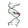
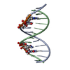
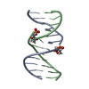
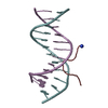
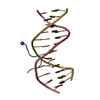
A: dodecamer DNA B: dodecamer DNA C: dodecamer DNA D: dodecamer DNA E: dodecamer DNA F: dodecamer DNA G: dodecamer DNA H: dodecamer DNA K: A-T hook peptide L: A-T hook peptide M: A-T hook peptide N: A-T hook peptide
Monochromator: mirrors / Protocol: SINGLE WAVELENGTH / Monochromatic (M) / Laue (L): M / Scattering type: x-ray
Radiation wavelength
Wavelength: 0.9795 Å / Relative weight: 1
Reflection
Resolution: 2→40 Å / Num. obs: 19462 / % possible obs: 94.3 %
Reflection shell
Resolution (Å)
Diffraction-ID
% possible all
2-2.03
1
56.4
2.03-2.07
1
70.1
2.07-2.11
1
86.8
2.11-2.15
1
93.7
2.15-2.2
1
97.2
2.2-2.25
1
97.7
2.25-2.31
1
98.2
2.31-2.37
1
98.5
2.37-2.44
1
98.1
2.44-2.52
1
98.7
2.52-2.61
1
98.5
2.61-2.71
1
98.6
2.71-2.84
1
98.8
2.84-2.99
1
99
2.99-3.17
1
98.8
3.17-3.42
1
98.6
3.42-3.76
1
99.3
3.76-4.31
1
99.4
4.31-5.42
1
99.5
5.42-40
1
98.3
-
Processing
Software
Name
Version
Classification
ADSC
Quantum
datacollection
AMoRE
phasing
REFMAC
5.6.0117
refinement
HKL-2000
datareduction
HKL-2000
datascaling
Refinement
Method to determine structure: MOLECULAR REPLACEMENT / Resolution: 2.27→37.76 Å / Cor.coef. Fo:Fc: 0.961 / Cor.coef. Fo:Fc free: 0.929 / SU B: 7.441 / SU ML: 0.184 / Cross valid method: THROUGHOUT / ESU R: 0.385 / ESU R Free: 0.268 / Stereochemistry target values: MAXIMUM LIKELIHOOD / Details: HYDROGENS HAVE BEEN USED IF PRESENT IN THE INPUT
Rfactor
Num. reflection
% reflection
Selection details
Rfree
0.2771
740
5.3 %
RANDOM
Rwork
0.21949
-
-
-
obs
0.22266
13163
98.76 %
-
all
-
19462
-
-
Solvent computation
Ion probe radii: 0.8 Å / Shrinkage radii: 0.8 Å / VDW probe radii: 1.2 Å / Solvent model: MASK
Displacement parameters
Biso mean: 35.433 Å2
Baniso -1
Baniso -2
Baniso -3
1-
3.33 Å2
0 Å2
0.9 Å2
2-
-
-3.85 Å2
0 Å2
3-
-
-
1.18 Å2
Refinement step
Cycle: LAST / Resolution: 2.27→37.76 Å
Protein
Nucleic acid
Ligand
Solvent
Total
Num. atoms
294
1944
0
59
2297
Refine LS restraints
Refine-ID
Type
Dev ideal
Dev ideal target
Number
X-RAY DIFFRACTION
r_bond_refined_d
0.009
0.012
2461
X-RAY DIFFRACTION
r_angle_refined_deg
2.221
1.437
3710
X-RAY DIFFRACTION
r_dihedral_angle_1_deg
7.964
5
30
X-RAY DIFFRACTION
r_dihedral_angle_2_deg
0.67
10
9
X-RAY DIFFRACTION
r_dihedral_angle_3_deg
23.246
15
72
X-RAY DIFFRACTION
r_dihedral_angle_4_deg
10.698
15
9
X-RAY DIFFRACTION
r_chiral_restr
0.103
0.2
318
X-RAY DIFFRACTION
r_gen_planes_refined
0.014
0.021
1205
LS refinement shell
Resolution: 2.27→2.329 Å / Total num. of bins used: 20
Rfactor
Num. reflection
% reflection
Rfree
0.387
47
-
Rwork
0.321
947
-
obs
-
-
98.12 %
+
About Yorodumi
-
News
-
Feb 9, 2022. New format data for meta-information of EMDB entries
New format data for meta-information of EMDB entries
Version 3 of the EMDB header file is now the official format.
The previous official version 1.9 will be removed from the archive.
In the structure databanks used in Yorodumi, some data are registered as the other names, "COVID-19 virus" and "2019-nCoV". Here are the details of the virus and the list of structure data.
Jan 31, 2019. EMDB accession codes are about to change! (news from PDBe EMDB page)
EMDB accession codes are about to change! (news from PDBe EMDB page)
The allocation of 4 digits for EMDB accession codes will soon come to an end. Whilst these codes will remain in use, new EMDB accession codes will include an additional digit and will expand incrementally as the available range of codes is exhausted. The current 4-digit format prefixed with “EMD-” (i.e. EMD-XXXX) will advance to a 5-digit format (i.e. EMD-XXXXX), and so on. It is currently estimated that the 4-digit codes will be depleted around Spring 2019, at which point the 5-digit format will come into force.
The EM Navigator/Yorodumi systems omit the EMD- prefix.
Related info.:Q: What is EMD? / ID/Accession-code notation in Yorodumi/EM Navigator
Yorodumi is a browser for structure data from EMDB, PDB, SASBDB, etc.
This page is also the successor to EM Navigator detail page, and also detail information page/front-end page for Omokage search.
The word "yorodu" (or yorozu) is an old Japanese word meaning "ten thousand". "mi" (miru) is to see.
Related info.:EMDB / PDB / SASBDB / Comparison of 3 databanks / Yorodumi Search / Aug 31, 2016. New EM Navigator & Yorodumi / Yorodumi Papers / Jmol/JSmol / Function and homology information / Changes in new EM Navigator and Yorodumi
 Movie
Movie Controller


 Open data
 Basic information
 Components
 Keywords
 Function and homology information
 X-RAY DIFFRACTION /
  Authors
 Citation
 Structure visualization
 Downloads & links
 Other downloads
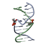


 PDBj
PDBj


 Assembly
 Mass: 18.015 Da / Num. of mol.: 59 / Source method: isolated from a natural source / Formula: H2O
 Sample preparation
 / Beamline: BM16 / Wavelength: 0.9795 Å
 Processing