 Movie
Movie Controller

[English] 日本語
 Yorodumi
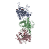
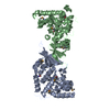
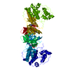
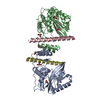
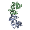
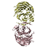
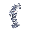
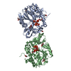
- PDB-1bg6: CRYSTAL STRUCTURE OF THE N-(1-D-CARBOXYLETHYL)-L-NORVALINE DEHYDR... -
+ Open data

- Basic information
| Entry | Database: PDB / ID: 1bg6 | ||||||
|---|---|---|---|---|---|---|---|
| Title | CRYSTAL STRUCTURE OF THE N-(1-D-CARBOXYLETHYL)-L-NORVALINE DEHYDROGENASE FROM ARTHROBACTER SP. STRAIN 1C | ||||||
 Components Components | N-(1-D-CARBOXYLETHYL)-L-NORVALINE DEHYDROGENASE | ||||||
 Keywords Keywords | OXIDOREDUCTASE / (D / L) STEREOSPECIFIC OPINE DEHYDROGENASE | ||||||
| Function / homology |  Function and homology information Function and homology information | ||||||
| Biological species |  Arthrobacter sp. (bacteria) Arthrobacter sp. (bacteria) | ||||||
| Method |  X-RAY DIFFRACTION / SYNCHROTRON / MIR / Resolution: 1.8 Å X-RAY DIFFRACTION / SYNCHROTRON / MIR / Resolution: 1.8 Å | ||||||
 Authors Authors | Britton, K.L. / Asano, Y. / Rice, D.W. | ||||||
 Citation Citation | Journal: Nat.Struct.Biol. / Year: 1998 Title: Crystal structure and active site location of N-(1-D-carboxylethyl)-L-norvaline dehydrogenase. Authors: Britton, K.L. / Asano, Y. / Rice, D.W. #1: Journal: Acta Crystallogr.,Sect.D / Year: 1998Title: Crystallization of Arthrobacter Sp. Strain 1C N-(1-D-Carboxyethyl)-L-Norvaline Dehydrogenase and its Complex with Nad+ Authors: Britton, K.L. / Rogers, H.F. / Asano, Y. / Dairi, T. / Kato, Y. / Stillman, T.J. / Rice, D.W. | ||||||
| History |
|
- Structure visualization
| Structure viewer | Molecule: MolmilJmol/JSmol |
|---|
- Downloads & links
-Download
| PDBx/mmCIF format | 1bg6.cif.gz | 77.9 KB | Display | PDBx/mmCIF format |
|---|---|---|---|---|
| PDB format | pdb1bg6.ent.gz | 58.2 KB | Display | PDB format |
| PDBx/mmJSON format | 1bg6.json.gz | Tree view | PDBx/mmJSON format | |
| Others |  Other downloads Other downloads |
-Validation report
| Arichive directory | https://data.pdbj.org/pub/pdb/validation_reports/bg/1bg6ftp://data.pdbj.org/pub/pdb/validation_reports/bg/1bg6 | HTTPS FTP |
|---|
-Related structure data
| Similar structure data |
|---|


-Links
 PDBj
PDBj


- Assembly
| Deposited unit | 
| ||||||||
|---|---|---|---|---|---|---|---|---|---|
| 1 | 
| ||||||||
| Unit cell |
|
-Components
| #1: Protein | Mass: 37969.188 Da / Num. of mol.: 1 Source method: isolated from a genetically manipulated source Source: (gene. exp.) Arthrobacter sp. (bacteria) / Strain: 1C / Gene: ODH / Plasmid: PODH1 / Production host: |
|---|---|
| #2: Water | ChemComp-HOH /  Mass: 18.015 Da / Num. of mol.: 113 / Source method: isolated from a natural source / Formula: H2O Mass: 18.015 Da / Num. of mol.: 113 / Source method: isolated from a natural source / Formula: H2O |
-Experimental details
-Experiment
| Experiment | Method: X-RAY DIFFRACTION / Number of used crystals: 2 |
|---|
- Sample preparation
| Crystal | Density Matthews: 2.65 Å3/Da / Density % sol: 41 % | |||||||||||||||||||||||||
|---|---|---|---|---|---|---|---|---|---|---|---|---|---|---|---|---|---|---|---|---|---|---|---|---|---|---|
| Crystal grow | pH: 6 / Details: pH 6.0 | |||||||||||||||||||||||||
| Crystal grow | *PLUS Temperature: 290 K / Method: vapor diffusion, hanging drop | |||||||||||||||||||||||||
| Components of the solutions | *PLUS
|
-Data collection
| Diffraction | Mean temperature: 290 K |
|---|---|
| Diffraction source | Source: SYNCHROTRON / Site: SRS  / Beamline: PX9.5 / Wavelength: 0.9 / Beamline: PX9.5 / Wavelength: 0.9 |
| Detector | Type: MARRESEARCH / Detector: IMAGE PLATE / Date: Dec 1, 1995 / Details: MIRRORS |
| Radiation | Monochromatic (M) / Laue (L): M / Scattering type: x-ray |
| Radiation wavelength | Wavelength: 0.9 Å / Relative weight: 1 |
| Reflection | Resolution: 1.8→16 Å / Num. obs: 30854 / % possible obs: 89 % / Observed criterion σ(I): 3 / Redundancy: 2.3 % / Rmerge(I) obs: 0.04 / Net I/σ(I): 8 |
| Reflection shell | Resolution: 1.8→1.85 Å / Redundancy: 2.4 % / Rmerge(I) obs: 0.152 / Mean I/σ(I) obs: 5 / % possible all: 78 |
| Reflection | *PLUS Num. measured all: 66518 |
| Reflection shell | *PLUS % possible obs: 78 % |
- Processing
| Software |
| ||||||||||||||||||||||||||||||
|---|---|---|---|---|---|---|---|---|---|---|---|---|---|---|---|---|---|---|---|---|---|---|---|---|---|---|---|---|---|---|---|
| Refinement | Method to determine structure: MIR / Resolution: 1.8→20 Å / σ(F): 0 / Stereochemistry target values: TNT PROTGEO /
| ||||||||||||||||||||||||||||||
| Solvent computation | Bsol: 310 Å2 / ksol: 0.85 e/Å3 | ||||||||||||||||||||||||||||||
| Refinement step | Cycle: LAST / Resolution: 1.8→20 Å
| ||||||||||||||||||||||||||||||
| Refine LS restraints |
| ||||||||||||||||||||||||||||||
| Software | *PLUS Name: TNT / Version: 5E / Classification: refinement | ||||||||||||||||||||||||||||||
| Refinement | *PLUS Num. reflection Rfree: 2545 / % reflection Rfree: 8 % / Rfactor obs: 0.188 / Rfactor Rfree: 0.263 | ||||||||||||||||||||||||||||||
| Solvent computation | *PLUS | ||||||||||||||||||||||||||||||
| Displacement parameters | *PLUS | ||||||||||||||||||||||||||||||
| Refine LS restraints | *PLUS
|
