 Movie
Movie Controller

[English] 日本語
 Yorodumi
- PDB-1b9y: STRUCTURAL ANALYSIS OF PHOSDUCIN AND ITS PHOSPHORYLATION-REGULATE... -
+ Open data

- Basic information
| Entry | Database: PDB / ID: 1b9y | ||||||
|---|---|---|---|---|---|---|---|
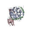
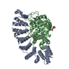
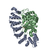
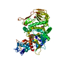
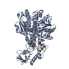
| Title | STRUCTURAL ANALYSIS OF PHOSDUCIN AND ITS PHOSPHORYLATION-REGULATED INTERACTION WITH TRANSDUCIN BETA-GAMMA | ||||||
 Components Components |
| ||||||
 Keywords Keywords | SIGNALING PROTEIN / PHOSDUCIN / TRANSDUCIN / BETA-GAMMA / SIGNAL TRANSDUCTION / REGULATION / PHOSPHORYLATION / G PROTEINS / THIOREDOXIN / VISION / MEKA / COMPLEX (TRANSDUCER- TRANSDUCTION) | ||||||
| Function / homology |  Function and homology information Function and homology informationOlfactory Signaling Pathway / Sensory perception of sweet, bitter, and umami (glutamate) taste / Synthesis, secretion, and inactivation of Glucagon-like Peptide-1 (GLP-1) / eye photoreceptor cell development / Inactivation, recovery and regulation of the phototransduction cascade / Activation of the phototransduction cascade / regulation of G protein-coupled receptor signaling pathway / Activation of G protein gated Potassium channels / G-protein activation / G beta:gamma signalling through PI3Kgamma ...Olfactory Signaling Pathway / Sensory perception of sweet, bitter, and umami (glutamate) taste / Synthesis, secretion, and inactivation of Glucagon-like Peptide-1 (GLP-1) / eye photoreceptor cell development / Inactivation, recovery and regulation of the phototransduction cascade / Activation of the phototransduction cascade / regulation of G protein-coupled receptor signaling pathway / Activation of G protein gated Potassium channels / G-protein activation / G beta:gamma signalling through PI3Kgamma / Prostacyclin signalling through prostacyclin receptor / G beta:gamma signalling through PLC beta / ADP signalling through P2Y purinoceptor 1 / Thromboxane signalling through TP receptor / Presynaptic function of Kainate receptors / G beta:gamma signalling through CDC42 / Inhibition of voltage gated Ca2+ channels via Gbeta/gamma subunits / G alpha (12/13) signalling events / Glucagon-type ligand receptors / G beta:gamma signalling through BTK / ADP signalling through P2Y purinoceptor 12 / Adrenaline,noradrenaline inhibits insulin secretion / Cooperation of PDCL (PhLP1) and TRiC/CCT in G-protein beta folding / Ca2+ pathway / G alpha (z) signalling events / Thrombin signalling through proteinase activated receptors (PARs) / Extra-nuclear estrogen signaling / G alpha (s) signalling events / G alpha (q) signalling events / G alpha (i) signalling events / Glucagon-like Peptide-1 (GLP1) regulates insulin secretion / High laminar flow shear stress activates signaling by PIEZO1 and PECAM1:CDH5:KDR in endothelial cells / Vasopressin regulates renal water homeostasis via Aquaporins / phototransduction / photoreceptor outer segment / photoreceptor inner segment / visual perception / photoreceptor disc membrane / intracellular protein localization / cellular response to catecholamine stimulus / adenylate cyclase-activating dopamine receptor signaling pathway / cellular response to prostaglandin E stimulus / heterotrimeric G-protein complex / G-protein beta-subunit binding / sensory perception of taste / signaling receptor complex adaptor activity / retina development in camera-type eye / GTPase binding / phospholipase C-activating G protein-coupled receptor signaling pathway / cell population proliferation / G protein-coupled receptor signaling pathway / GTPase activity / synapse / protein-containing complex binding / membrane / nucleus / cytoplasm / cytosol Similarity search - Function | ||||||
| Biological species |  | ||||||
| Method |  X-RAY DIFFRACTION / SYNCHROTRON / MOLECULAR REPLACEMENT / Resolution: 3 Å X-RAY DIFFRACTION / SYNCHROTRON / MOLECULAR REPLACEMENT / Resolution: 3 Å | ||||||
 Authors Authors | Gaudet, R. / Sigler, P.B. | ||||||
 Citation Citation | Journal: Mol.Cell / Year: 1999 Title: A molecular mechanism for the phosphorylation-dependent regulation of heterotrimeric G proteins by phosducin. Authors: Gaudet, R. / Savage, J.R. / McLaughlin, J.N. / Willardson, B.M. / Sigler, P.B. #1: Journal: Nature / Year: 1996Title: Crystal structure of a G-protein beta gamma dimer at 2.1A resolution. Authors: Sondek, J. / Bohm, A. / Lambright, D.G. / Hamm, H.E. / Sigler, P.B. #2: Journal: Cell(Cambridge,Mass.) / Year: 1996Title: Crystal structure at 2.4 angstroms resolution of the complex of transducin betagamma and its regulator, phosducin. Authors: Gaudet, R. / Bohm, A. / Sigler, P.B. | ||||||
| History |
|
- Structure visualization
| Structure viewer | Molecule: MolmilJmol/JSmol |
|---|
- Downloads & links
-Download
| PDBx/mmCIF format | 1b9y.cif.gz | 119.1 KB | Display | PDBx/mmCIF format |
|---|---|---|---|---|
| PDB format | pdb1b9y.ent.gz | 91.6 KB | Display | PDB format |
| PDBx/mmJSON format | 1b9y.json.gz | Tree view | PDBx/mmJSON format | |
| Others |  Other downloads Other downloads |
-Validation report
| Arichive directory | https://data.pdbj.org/pub/pdb/validation_reports/b9/1b9yftp://data.pdbj.org/pub/pdb/validation_reports/b9/1b9y | HTTPS FTP |
|---|
-Related structure data
| Related structure data |  1b9xC 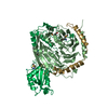 2trcS S: Starting model for refinement C: citing same article ( |
|---|---|
| Similar structure data |

-Links
 PDBj
PDBj


- Assembly
| Deposited unit | 
| ||||||||||
|---|---|---|---|---|---|---|---|---|---|---|---|
| 1 |
| ||||||||||
| Unit cell |
|
-Components
| #1: Protein | Mass: 37430.957 Da / Num. of mol.: 1 / Fragment: LYS-C RESISTANT FRAGMENT, THE BETA SUBUNIT / Source method: isolated from a natural source / Details: PURIFIED FROM BOVINE ROD OUTER SEGMENTS / Source: (natural) | ||||
|---|---|---|---|---|---|
| #2: Protein | Mass: 8040.304 Da / Num. of mol.: 1 Fragment: LYS-C RESISTANT FRAGMENT, THE GAMMA SUBUNIT CLEAVED AFTER RESIDUE 68 Source method: isolated from a natural source / Details: PURIFIED FROM BOVINE ROD OUTER SEGMENTS / Source: (natural) | ||||
| #3: Protein | Mass: 28162.273 Da / Num. of mol.: 1 Source method: isolated from a genetically manipulated source Source: (gene. exp.) Description: N-TERMINAL EXTENSION OF THE SEQUENCE MGSSHHHHHHSSGLVPRGSH. Cellular location: CYTOSOLIC / Gene: RAT PDC / Organ: PINEAL GLAND, RETINA / Plasmid: PET15B/PHOSDUCIN / Species (production host): Escherichia coli / Gene (production host): RAT PDC / Production host:  | ||||
| #4: Chemical | ChemComp-GD /   Mass: 157.250 Da / Num. of mol.: 6 / Source method: obtained synthetically / Formula: Gd Mass: 157.250 Da / Num. of mol.: 6 / Source method: obtained synthetically / Formula: Gd#5: Water | ChemComp-HOH / |  Mass: 18.015 Da / Num. of mol.: 20 / Source method: isolated from a natural source / Formula: H2O Mass: 18.015 Da / Num. of mol.: 20 / Source method: isolated from a natural source / Formula: H2OCompound details | PHOSPHORYLATION SITE S73 IN PHOSDUCIN IS PHOSPHORYLATED IN THIS STRUCTURE BUT IS NOT VISIBLE IN THE ...PHOSPHORYL | |
-Experimental details
-Experiment
| Experiment | Method: X-RAY DIFFRACTION / Number of used crystals: 1 |
|---|
- Sample preparation
| Crystal | Density Matthews: 2.7 Å3/Da / Density % sol: 47 % | |||||||||||||||||||||||||||||||||||||||||||||
|---|---|---|---|---|---|---|---|---|---|---|---|---|---|---|---|---|---|---|---|---|---|---|---|---|---|---|---|---|---|---|---|---|---|---|---|---|---|---|---|---|---|---|---|---|---|---|
| Crystal grow | Temperature: 277 K / Method: vapor diffusion, hanging drop / pH: 5.5 Details: pH 5.5, VAPOR DIFFUSION, HANGING DROP, temperature 277K | |||||||||||||||||||||||||||||||||||||||||||||
| Components of the solutions |
| |||||||||||||||||||||||||||||||||||||||||||||
| Crystal grow | *PLUS Temperature: 4 ℃ | |||||||||||||||||||||||||||||||||||||||||||||
| Components of the solutions | *PLUS
|
-Data collection
| Diffraction | Mean temperature: 110 K |
|---|---|
| Diffraction source | Source: SYNCHROTRON / Site: CHESS  / Beamline: A1 / Wavelength: 0.92 / Beamline: A1 / Wavelength: 0.92 |
| Detector | Type: PRINCETON 2K / Detector: CCD / Date: Aug 15, 1998 / Details: MIRRORS |
| Radiation | Protocol: SINGLE WAVELENGTH / Monochromatic (M) / Laue (L): M / Scattering type: x-ray |
| Radiation wavelength | Wavelength: 0.92 Å / Relative weight: 1 |
| Reflection | Resolution: 3→50 Å / Num. all: 13650 / Num. obs: 13650 / % possible obs: 94.5 % / Observed criterion σ(I): 0 / Redundancy: 3.1 % / Rsym value: 0.085 / Net I/σ(I): 10.7 |
| Reflection shell | Resolution: 3→3.09 Å / Mean I/σ(I) obs: 2.9 / Rsym value: 0.356 / % possible all: 97.1 |
| Reflection | *PLUS % possible obs: 94.5 % / Redundancy: 3.1 % / Rmerge(I) obs: 0.085 |
| Reflection shell | *PLUS % possible obs: 97.1 % / Rmerge(I) obs: 0.356 / Mean I/σ(I) obs: 2.9 |
- Processing
| Software |
| ||||||||||||||||||||||||||||||||||||||||||||||||||||||||||||
|---|---|---|---|---|---|---|---|---|---|---|---|---|---|---|---|---|---|---|---|---|---|---|---|---|---|---|---|---|---|---|---|---|---|---|---|---|---|---|---|---|---|---|---|---|---|---|---|---|---|---|---|---|---|---|---|---|---|---|---|---|---|
| Refinement | Method to determine structure: MOLECULAR REPLACEMENT Starting model: PDB ENTRY 2TRC Resolution: 3→50 Å / Rfactor Rfree error: 0.008 / Data cutoff high rms absF: 329567.14 / Isotropic thermal model: GROUP / Cross valid method: THROUGHOUT / σ(F): 2 Details: DISORDERED REGION IN PHOSDUCIN FROM RESIDUE 37 - 38 WAS MODELED STEREOCHEMICALLY AS A POLYALANINE CHAIN. DISORDERED REGION IN PHOSDUCIN FROM RESIDUE 39 - 86 WAS NOT VISIBLE IN THE MAPS AND WAS NOT MODELLED
| ||||||||||||||||||||||||||||||||||||||||||||||||||||||||||||
| Solvent computation | Solvent model: FLAT MODEL / Bsol: 34.58 Å2 / ksol: 0.313 e/Å3 | ||||||||||||||||||||||||||||||||||||||||||||||||||||||||||||
| Displacement parameters | Biso mean: 67.9 Å2
| ||||||||||||||||||||||||||||||||||||||||||||||||||||||||||||
| Refine analyze |
| ||||||||||||||||||||||||||||||||||||||||||||||||||||||||||||
| Refinement step | Cycle: LAST / Resolution: 3→50 Å
| ||||||||||||||||||||||||||||||||||||||||||||||||||||||||||||
| Refine LS restraints |
| ||||||||||||||||||||||||||||||||||||||||||||||||||||||||||||
| LS refinement shell | Resolution: 3→3.19 Å / Rfactor Rfree error: 0.029 / Total num. of bins used: 6
| ||||||||||||||||||||||||||||||||||||||||||||||||||||||||||||
| Xplor file |
| ||||||||||||||||||||||||||||||||||||||||||||||||||||||||||||
| Software | *PLUS Name: CNS / Version: 0.5 / Classification: refinement | ||||||||||||||||||||||||||||||||||||||||||||||||||||||||||||
| Refinement | *PLUS Num. reflection all: 13650 / σ(F): 2 / % reflection Rfree: 9.7 % / Rfactor all: 0.224 / Rfactor obs: 0.213 | ||||||||||||||||||||||||||||||||||||||||||||||||||||||||||||
| Solvent computation | *PLUS | ||||||||||||||||||||||||||||||||||||||||||||||||||||||||||||
| Displacement parameters | *PLUS Biso mean: 67.9 Å2 | ||||||||||||||||||||||||||||||||||||||||||||||||||||||||||||
| Refine LS restraints | *PLUS
| ||||||||||||||||||||||||||||||||||||||||||||||||||||||||||||
| LS refinement shell | *PLUS Rfactor Rfree: 0.37 / % reflection Rfree: 9.3 % / Rfactor Rwork: 0.288 / Num. reflection obs: 1855 / Rfactor all: 0.33 / Rfactor obs: 0.288 |
