 Movie
Movie Controller


+ Open data

- Basic information
| Entry | Database: PDB / ID: 2trc | ||||||
|---|---|---|---|---|---|---|---|
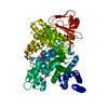
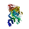
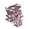
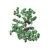
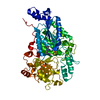
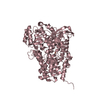
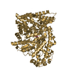
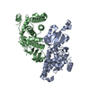
| Title | PHOSDUCIN/TRANSDUCIN BETA-GAMMA COMPLEX | ||||||
 Components Components |
| ||||||
 Keywords Keywords | COMPLEX (TRANSDUCER/TRANSDUCTION) / PHOSDUCIN / TRANSDUCIN / BETA-GAMMA / SIGNAL TRANSDUCTION / REGULATION / PHOSPHORYLATION / G PROTEINS / THIOREDOXIN / VISION / MEKA / COMPLEX (TRANSDUCER-TRANSDUCTION) / COMPLEX (TRANSDUCER-TRANSDUCTION) complex | ||||||
| Function / homology |  Function and homology information Function and homology informationOlfactory Signaling Pathway / Sensory perception of sweet, bitter, and umami (glutamate) taste / Synthesis, secretion, and inactivation of Glucagon-like Peptide-1 (GLP-1) / eye photoreceptor cell development / Inactivation, recovery and regulation of the phototransduction cascade / Activation of the phototransduction cascade / regulation of G protein-coupled receptor signaling pathway / Activation of G protein gated Potassium channels / G-protein activation / G beta:gamma signalling through PI3Kgamma ...Olfactory Signaling Pathway / Sensory perception of sweet, bitter, and umami (glutamate) taste / Synthesis, secretion, and inactivation of Glucagon-like Peptide-1 (GLP-1) / eye photoreceptor cell development / Inactivation, recovery and regulation of the phototransduction cascade / Activation of the phototransduction cascade / regulation of G protein-coupled receptor signaling pathway / Activation of G protein gated Potassium channels / G-protein activation / G beta:gamma signalling through PI3Kgamma / Prostacyclin signalling through prostacyclin receptor / G beta:gamma signalling through PLC beta / ADP signalling through P2Y purinoceptor 1 / Thromboxane signalling through TP receptor / Presynaptic function of Kainate receptors / G beta:gamma signalling through CDC42 / Inhibition of voltage gated Ca2+ channels via Gbeta/gamma subunits / G alpha (12/13) signalling events / Glucagon-type ligand receptors / G beta:gamma signalling through BTK / ADP signalling through P2Y purinoceptor 12 / Adrenaline,noradrenaline inhibits insulin secretion / Cooperation of PDCL (PhLP1) and TRiC/CCT in G-protein beta folding / Ca2+ pathway / G alpha (z) signalling events / Thrombin signalling through proteinase activated receptors (PARs) / Extra-nuclear estrogen signaling / G alpha (s) signalling events / G alpha (q) signalling events / G alpha (i) signalling events / Glucagon-like Peptide-1 (GLP1) regulates insulin secretion / High laminar flow shear stress activates signaling by PIEZO1 and PECAM1:CDH5:KDR in endothelial cells / Vasopressin regulates renal water homeostasis via Aquaporins / phototransduction / photoreceptor outer segment / photoreceptor inner segment / visual perception / photoreceptor disc membrane / intracellular protein localization / cellular response to catecholamine stimulus / adenylate cyclase-activating dopamine receptor signaling pathway / cellular response to prostaglandin E stimulus / heterotrimeric G-protein complex / G-protein beta-subunit binding / sensory perception of taste / signaling receptor complex adaptor activity / retina development in camera-type eye / GTPase binding / phospholipase C-activating G protein-coupled receptor signaling pathway / cell population proliferation / G protein-coupled receptor signaling pathway / GTPase activity / synapse / protein-containing complex binding / membrane / nucleus / cytoplasm / cytosol Similarity search - Function | ||||||
| Biological species |  | ||||||
| Method |  X-RAY DIFFRACTION / SYNCHROTRON / MAD / Resolution: 2.4 Å X-RAY DIFFRACTION / SYNCHROTRON / MAD / Resolution: 2.4 Å | ||||||
 Authors Authors | Gaudet, R. / Bohm, A. / Sigler, P.B. | ||||||
 Citation Citation | Journal: Cell(Cambridge,Mass.) / Year: 1996 Title: Crystal structure at 2.4 angstroms resolution of the complex of transducin betagamma and its regulator, phosducin. Authors: Gaudet, R. / Bohm, A. / Sigler, P.B. #1: Journal: Nature / Year: 1996Title: Crystal Structure of a Ga Protein Beta Gamma Dimer at 2.1A Resolution Authors: Sondek, J. / Bohm, A. / Lambright, D.G. / Hamm, H.E. / Sigler, P.B. | ||||||
| History |
|
- Structure visualization
| Structure viewer | Molecule: MolmilJmol/JSmol |
|---|
- Downloads & links
-Download
| PDBx/mmCIF format | 2trc.cif.gz | 139.9 KB | Display | PDBx/mmCIF format |
|---|---|---|---|---|
| PDB format | pdb2trc.ent.gz | 107.5 KB | Display | PDB format |
| PDBx/mmJSON format | 2trc.json.gz | Tree view | PDBx/mmJSON format | |
| Others |  Other downloads Other downloads |
-Validation report
| Arichive directory | https://data.pdbj.org/pub/pdb/validation_reports/tr/2trcftp://data.pdbj.org/pub/pdb/validation_reports/tr/2trc | HTTPS FTP |
|---|
-Related structure data
| Similar structure data |
|---|


-Links
 PDBj
PDBj


- Assembly
| Deposited unit | 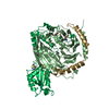
| ||||||||
|---|---|---|---|---|---|---|---|---|---|
| 1 |
| ||||||||
| Unit cell |
|
-Components
| #1: Protein | Mass: 37430.957 Da / Num. of mol.: 1 Fragment: LYS-C RESISTANT FRAGMENT, THE GAMMA SUBUNIT CLEAVED AFTER RESIDUE 68 Source method: isolated from a natural source / Details: PURIFIED FROM BOVINE ROD OUTER SEGMENTS / Source: (natural) | ||||
|---|---|---|---|---|---|
| #2: Protein | Mass: 8040.304 Da / Num. of mol.: 1 Fragment: LYS-C RESISTANT FRAGMENT, THE GAMMA SUBUNIT CLEAVED AFTER RESIDUE 68 Source method: isolated from a natural source / Details: PURIFIED FROM BOVINE ROD OUTER SEGMENTS / Source: (natural) | ||||
| #3: Protein | Mass: 25088.402 Da / Num. of mol.: 1 Source method: isolated from a genetically manipulated source Source: (gene. exp.) | ||||
| #4: Chemical | ChemComp-GD /   Mass: 157.250 Da / Num. of mol.: 5 / Source method: obtained synthetically / Formula: Gd Mass: 157.250 Da / Num. of mol.: 5 / Source method: obtained synthetically / Formula: Gd#5: Water | ChemComp-HOH / |  Mass: 18.015 Da / Num. of mol.: 234 / Source method: isolated from a natural source / Formula: H2O Mass: 18.015 Da / Num. of mol.: 234 / Source method: isolated from a natural source / Formula: H2OHas protein modification | Y | |
-Experimental details
-Experiment
| Experiment | Method: X-RAY DIFFRACTION / Number of used crystals: 1 |
|---|
- Sample preparation
| Crystal | Density Matthews: 2.7 Å3/Da / Density % sol: 47 % | |||||||||||||||||||||||||||||||||||||||||||||
|---|---|---|---|---|---|---|---|---|---|---|---|---|---|---|---|---|---|---|---|---|---|---|---|---|---|---|---|---|---|---|---|---|---|---|---|---|---|---|---|---|---|---|---|---|---|---|
| Crystal grow | Temperature: 277 K / Method: vapor diffusion, hanging drop / pH: 5 Details: THE PROTEIN COMPLEX (10 MG/ML SOLUTION) WAS CRYSTALLIZED FROM 50 MM SODIUM CITRATE (PH 5.0), 150 MM MAGNESIUM ACETATE, 9.5% PEG 8000, BY HANGING DROP METHOD AT 4 DEGREES C., vapor diffusion - ...Details: THE PROTEIN COMPLEX (10 MG/ML SOLUTION) WAS CRYSTALLIZED FROM 50 MM SODIUM CITRATE (PH 5.0), 150 MM MAGNESIUM ACETATE, 9.5% PEG 8000, BY HANGING DROP METHOD AT 4 DEGREES C., vapor diffusion - hanging drop, temperature 277K | |||||||||||||||||||||||||||||||||||||||||||||
| Crystal grow | *PLUS Temperature: 4 ℃ / Method: vapor diffusion, hanging drop | |||||||||||||||||||||||||||||||||||||||||||||
| Components of the solutions | *PLUS
|
-Data collection
| Diffraction | Mean temperature: 110 K | |||||||||
|---|---|---|---|---|---|---|---|---|---|---|
| Diffraction source | Source: SYNCHROTRON / Site: NSLS  / Beamline: X4A / Wavelength: 0.9791 / Wavelength: 0.9791, 1.7109 / Beamline: X4A / Wavelength: 0.9791 / Wavelength: 0.9791, 1.7109 | |||||||||
| Detector | Type: FUJI / Detector: IMAGE PLATE / Date: Jun 6, 1996 / Details: MIRRORS | |||||||||
| Radiation | Monochromator: SI(111) / Monochromatic (M) / Laue (L): M / Scattering type: x-ray | |||||||||
| Radiation wavelength |
| |||||||||
| Reflection | Resolution: 2.4→50 Å / Num. obs: 24085 / % possible obs: 94.9 % / Observed criterion σ(I): 2 / Redundancy: 3 % / Biso Wilson estimate: 37 Å2 / Rsym value: 0.06 / Net I/σ(I): 16 | |||||||||
| Reflection shell | Resolution: 2.37→2.44 Å / Redundancy: 3 % / Mean I/σ(I) obs: 2.9 / Rsym value: 0.301 / % possible all: 97 | |||||||||
| Reflection | *PLUS Rmerge(I) obs: 0.06 | |||||||||
| Reflection shell | *PLUS % possible obs: 96.8 % / Rmerge(I) obs: 0.301 |
- Processing
| Software |
| ||||||||||||||||||||||||||||||||||||||||||||||||||||||||||||||||||||||||||||||||
|---|---|---|---|---|---|---|---|---|---|---|---|---|---|---|---|---|---|---|---|---|---|---|---|---|---|---|---|---|---|---|---|---|---|---|---|---|---|---|---|---|---|---|---|---|---|---|---|---|---|---|---|---|---|---|---|---|---|---|---|---|---|---|---|---|---|---|---|---|---|---|---|---|---|---|---|---|---|---|---|---|---|
| Refinement | Method to determine structure: MAD / Resolution: 2.4→50 Å / Rfactor Rfree error: 0.0057 / Data cutoff high absF: 100000 / Data cutoff low absF: 0.1 / Isotropic thermal model: RESTRAINED / Cross valid method: THROUGHOUT / σ(F): 2 Details: DISORDERED REGION IN PHOSDUCIN FROM RESIDUE 37 - 66 WAS MODELED STEREOCHEMICALLY AS A POLYALANINE CHAIN.
| ||||||||||||||||||||||||||||||||||||||||||||||||||||||||||||||||||||||||||||||||
| Displacement parameters | Biso mean: 27.2 Å2
| ||||||||||||||||||||||||||||||||||||||||||||||||||||||||||||||||||||||||||||||||
| Refine analyze |
| ||||||||||||||||||||||||||||||||||||||||||||||||||||||||||||||||||||||||||||||||
| Refinement step | Cycle: LAST / Resolution: 2.4→50 Å
| ||||||||||||||||||||||||||||||||||||||||||||||||||||||||||||||||||||||||||||||||
| Refine LS restraints |
| ||||||||||||||||||||||||||||||||||||||||||||||||||||||||||||||||||||||||||||||||
| LS refinement shell | Resolution: 2.4→2.51 Å / Rfactor Rfree error: 0.019 / Total num. of bins used: 8
| ||||||||||||||||||||||||||||||||||||||||||||||||||||||||||||||||||||||||||||||||
| Xplor file |
| ||||||||||||||||||||||||||||||||||||||||||||||||||||||||||||||||||||||||||||||||
| Software | *PLUS Name: X-PLOR / Version: 3.8 / Classification: refinement | ||||||||||||||||||||||||||||||||||||||||||||||||||||||||||||||||||||||||||||||||
| Refinement | *PLUS Rfactor obs: 0.19 / Rfactor Rwork: 0.19 | ||||||||||||||||||||||||||||||||||||||||||||||||||||||||||||||||||||||||||||||||
| Solvent computation | *PLUS | ||||||||||||||||||||||||||||||||||||||||||||||||||||||||||||||||||||||||||||||||
| Displacement parameters | *PLUS | ||||||||||||||||||||||||||||||||||||||||||||||||||||||||||||||||||||||||||||||||
| Refine LS restraints | *PLUS
|
