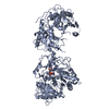
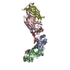
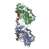
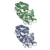
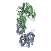
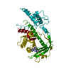
transposase activity / DNA transposition / endonuclease activity / Hydrolases; Acting on ester bonds / DNA binding / metal ion binding Similarity search - Function
Transferase Inhibitor Protein From Tn5; Chain A, domain2 / Transferase Inhibitor Protein From Tn5; Chain / Transposase Inhibitor Protein From Tn5; Chain A, domain 1 / Transposase Inhibitor Protein From Tn5; Chain A, domain 1 / Transposase, IS4-like / Transposase, Tn5-like, N-terminal / Transposase, Tn5-like, C-terminal / Transposase, Tn5-like, N-terminal domain superfamily / : / : ...Transferase Inhibitor Protein From Tn5; Chain A, domain2 / Transferase Inhibitor Protein From Tn5; Chain / Transposase Inhibitor Protein From Tn5; Chain A, domain 1 / Transposase Inhibitor Protein From Tn5; Chain A, domain 1 / Transposase, IS4-like / Transposase, Tn5-like, N-terminal / Transposase, Tn5-like, C-terminal / Transposase, Tn5-like, N-terminal domain superfamily / : / : / Transposase DDE domain / Transposase DNA-binding / Ribonuclease H-like superfamily / Alpha-Beta Complex / Orthogonal Bundle / Mainly Alpha / Alpha Beta Similarity search - Domain/homology
Mass: 18.015 Da / Num. of mol.: 56 / Source method: isolated from a natural source / Formula: H2O
Sequence details
THE FOLLOWING RESIDUES WERE DISORDERED BEYOND THE BETA-CARBON AND THUS WERE MODELED AS ALANINE: ...THE FOLLOWING RESIDUES WERE DISORDERED BEYOND THE BETA-CARBON AND THUS WERE MODELED AS ALANINE: GLU72, GLU88, ASP133, ARG215, LYS216, LYS244, VAL246, GLN341, ARG342, MET343, PRO346, ASP347, ASN348, MET352, ASP400, AND GLU 417 ALA 94 WAS DISORDERED BEYOND THE ALPHA-CARBON AND WAS MODELED AS GLYCINE.
-
Experimental details
-
Experiment
Experiment
Method: X-RAY DIFFRACTION / Number of used crystals: 1
-
Sample preparation
Crystal
Density Matthews: 2.84 Å3/Da / Density % sol: 57 % / Description: STRUCTURE WAS INDEPENDENTLY SOLVED USING MIR
In the structure databanks used in Yorodumi, some data are registered as the other names, "COVID-19 virus" and "2019-nCoV". Here are the details of the virus and the list of structure data.
Jan 31, 2019. EMDB accession codes are about to change! (news from PDBe EMDB page)
EMDB accession codes are about to change! (news from PDBe EMDB page)
The allocation of 4 digits for EMDB accession codes will soon come to an end. Whilst these codes will remain in use, new EMDB accession codes will include an additional digit and will expand incrementally as the available range of codes is exhausted. The current 4-digit format prefixed with “EMD-” (i.e. EMD-XXXX) will advance to a 5-digit format (i.e. EMD-XXXXX), and so on. It is currently estimated that the 4-digit codes will be depleted around Spring 2019, at which point the 5-digit format will come into force.
The EM Navigator/Yorodumi systems omit the EMD- prefix.
Related info.:Q: What is EMD? / ID/Accession-code notation in Yorodumi/EM Navigator
Yorodumi is a browser for structure data from EMDB, PDB, SASBDB, etc.
This page is also the successor to EM Navigator detail page, and also detail information page/front-end page for Omokage search.
The word "yorodu" (or yorozu) is an old Japanese word meaning "ten thousand". "mi" (miru) is to see.
Related info.:EMDB / PDB / SASBDB / Comparison of 3 databanks / Yorodumi Search / Aug 31, 2016. New EM Navigator & Yorodumi / Yorodumi Papers / Jmol/JSmol / Function and homology information / Changes in new EM Navigator and Yorodumi
 Movie
Movie Controller


 Open data
 Basic information
 Components
 Keywords
 Function and homology information
 X-RAY DIFFRACTION /
  Authors
 Citation
 Structure visualization
 Downloads & links
 Other downloads
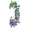
 PDBj
PDBj
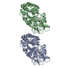
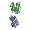
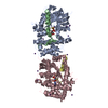
 Assembly
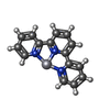
 Mass: 463.799 Da / Num. of mol.: 2 / Source method: obtained synthetically / Formula: C15H11ClN3Pt
 Mass: 18.015 Da / Num. of mol.: 56 / Source method: isolated from a natural source / Formula: H2O
 Sample preparation
 / Beamline: 19-ID / Wavelength: 1.0273,1.0720,1.1201
 Processing