 Movie
Movie Controller

[English] 日本語
 Yorodumi
- PDB-2v8d: Crystal structure of mutant E159A of beta-alanine synthase from S... -
+ Open data

- Basic information
| Entry | Database: PDB / ID: 2v8d | ||||||
|---|---|---|---|---|---|---|---|
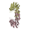
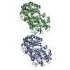
| Title | Crystal structure of mutant E159A of beta-alanine synthase from Saccharomyces kluyveri | ||||||
 Components Components | BETA-ALANINE SYNTHASE | ||||||
 Keywords Keywords | HYDROLASE / DI-ZINC CENTER / AMIDOHYDROLASE | ||||||
| Function / homology |  Function and homology information Function and homology informationbeta-ureidopropionase activity / beta-ureidopropionase / hydrolase activity, acting on carbon-nitrogen (but not peptide) bonds, in linear amidines / metal ion binding Similarity search - Function | ||||||
| Biological species |  SACCHAROMYCES KLUYVERI (fungus) SACCHAROMYCES KLUYVERI (fungus) | ||||||
| Method |  X-RAY DIFFRACTION / SYNCHROTRON / MOLECULAR REPLACEMENT / Resolution: 2.3 Å X-RAY DIFFRACTION / SYNCHROTRON / MOLECULAR REPLACEMENT / Resolution: 2.3 Å | ||||||
 Authors Authors | Lundgren, S. / Andersen, B. / Piskur, J. / Dobritzsch, D. | ||||||
 Citation Citation | Journal: J.Biol.Chem. / Year: 2007 Title: Crystal Structures of Yeast -Alanine Synthase Complexes Reveal the Mode of Substrate Binding and Large Scale Domain Closure Movements. Authors: Lundgren, S. / Andersen, B. / Piskur, J. / Dobritzsch, D. #1: Journal: J.Biol.Chem. / Year: 2003Title: Yeast Beta-Alanine Synthase Shares a Structural Scaffold and Origin with Dizinc-Dependent Exopeptidases Authors: Lundgren, S. / Gojkovic, Z. / Piskur, J. / Dobritzsch, D. #2: Journal: Acta Crystallogr.,Sect.D / Year: 2003 Title: Crystallization and Preliminary X-Ray Analysis of Beta-Alanine Synthase from the Yeast Saccharomyces Kluyveri Authors: Dobritzsch, D. / Gojkovic, Z. / Andersen, B. / Piskur, J. | ||||||
| History |
| ||||||
| Remark 700 | SHEET THE SHEET STRUCTURE OF THIS MOLECULE IS BIFURCATED. IN ORDER TO REPRESENT THIS FEATURE IN ... SHEET THE SHEET STRUCTURE OF THIS MOLECULE IS BIFURCATED. IN ORDER TO REPRESENT THIS FEATURE IN THE SHEET RECORDS BELOW, TWO SHEETS ARE DEFINED. |
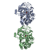
- Structure visualization
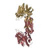
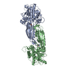
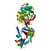
| Structure viewer | Molecule: MolmilJmol/JSmol |
|---|
- Downloads & links
-Download
| PDBx/mmCIF format | 2v8d.cif.gz | 176.5 KB | Display | PDBx/mmCIF format |
|---|---|---|---|---|
| PDB format | pdb2v8d.ent.gz | 139.4 KB | Display | PDB format |
| PDBx/mmJSON format | 2v8d.json.gz | Tree view | PDBx/mmJSON format | |
| Others |  Other downloads Other downloads |
-Validation report
| Arichive directory | https://data.pdbj.org/pub/pdb/validation_reports/v8/2v8dftp://data.pdbj.org/pub/pdb/validation_reports/v8/2v8d | HTTPS FTP |
|---|
-Related structure data
| Related structure data |  2v8gC 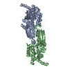 2v8hC 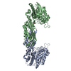 2v8vC  1r43S C: citing same article ( S: Starting model for refinement |
|---|---|
| Similar structure data |


-Links
 PDBj
PDBj
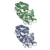
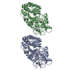
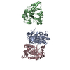
- Assembly
| Deposited unit | 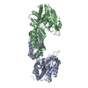
| |||||||||||||||||||||||||||||||||||||||||||||||||||||||||||||||||||
|---|---|---|---|---|---|---|---|---|---|---|---|---|---|---|---|---|---|---|---|---|---|---|---|---|---|---|---|---|---|---|---|---|---|---|---|---|---|---|---|---|---|---|---|---|---|---|---|---|---|---|---|---|---|---|---|---|---|---|---|---|---|---|---|---|---|---|---|---|
| 1 |
| |||||||||||||||||||||||||||||||||||||||||||||||||||||||||||||||||||
| Unit cell |
| |||||||||||||||||||||||||||||||||||||||||||||||||||||||||||||||||||
| Noncrystallographic symmetry (NCS) | NCS domain:
NCS domain segments:
NCS oper:
|
-Components
| #1: Protein | Mass: 51948.773 Da / Num. of mol.: 2 / Fragment: RESIDUES 2-455 / Mutation: YES Source method: isolated from a genetically manipulated source Source: (gene. exp.) SACCHAROMYCES KLUYVERI (fungus) / Production host:  #2: Chemical | ChemComp-ZN /   Mass: 65.409 Da / Num. of mol.: 4 / Source method: obtained synthetically / Formula: Zn Mass: 65.409 Da / Num. of mol.: 4 / Source method: obtained synthetically / Formula: Zn#3: Water | ChemComp-HOH / |  Mass: 18.015 Da / Num. of mol.: 58 / Source method: isolated from a natural source / Formula: H2O Mass: 18.015 Da / Num. of mol.: 58 / Source method: isolated from a natural source / Formula: H2OCompound details | ENGINEERED | Sequence details | THE N-TERMINAL METHIONINE IS ABSENT, MOST LIKELY DUE TO POSTTRANSLATIONAL MODIFICATION. THE LAST 20 ...THE N-TERMINAL METHIONINE | |
|---|
-Experimental details
-Experiment
| Experiment | Method: X-RAY DIFFRACTION / Number of used crystals: 1 |
|---|
- Sample preparation
| Crystal | Density Matthews: 2.45 Å3/Da / Density % sol: 50 % / Description: NONE |
|---|---|
| Crystal grow | Temperature: 293 K / Method: vapor diffusion, hanging drop / pH: 6 Details: TRISODIUM CITRATE, DIOXANE, ACETONE, DTT, PH 6.0, VAPOR DIFFUSION, HANGING DROP, TEMPERATURE 293K |
-Data collection
| Diffraction | Mean temperature: 100 K |
|---|---|
| Diffraction source | Source: SYNCHROTRON / Site: ESRF  / Beamline: ID14-4 / Wavelength: 0.9394 / Beamline: ID14-4 / Wavelength: 0.9394 |
| Detector | Type: ADSC CCD / Detector: CCD / Date: May 7, 2006 |
| Radiation | Protocol: SINGLE WAVELENGTH / Monochromatic (M) / Laue (L): M / Scattering type: x-ray |
| Radiation wavelength | Wavelength: 0.9394 Å / Relative weight: 1 |
| Reflection | Resolution: 2.3→50 Å / Num. obs: 43156 / % possible obs: 97 % / Observed criterion σ(I): 2 / Redundancy: 3.2 % / Biso Wilson estimate: 48.8 Å2 / Rmerge(I) obs: 0.08 / Net I/σ(I): 11.9 |
| Reflection shell | Resolution: 2.3→2.4 Å / Redundancy: 2.7 % / Rmerge(I) obs: 0.46 / Mean I/σ(I) obs: 2 / % possible all: 88 |
- Processing
| Software |
| ||||||||||||||||||||||||||||||||||||||||||||||||||||||||||||||||||||||||||||||||||||||||||||||||||||||||||||||||||||||||||||||||||||||||||||||||||||||||||||||||||||||||||||||||||||||
|---|---|---|---|---|---|---|---|---|---|---|---|---|---|---|---|---|---|---|---|---|---|---|---|---|---|---|---|---|---|---|---|---|---|---|---|---|---|---|---|---|---|---|---|---|---|---|---|---|---|---|---|---|---|---|---|---|---|---|---|---|---|---|---|---|---|---|---|---|---|---|---|---|---|---|---|---|---|---|---|---|---|---|---|---|---|---|---|---|---|---|---|---|---|---|---|---|---|---|---|---|---|---|---|---|---|---|---|---|---|---|---|---|---|---|---|---|---|---|---|---|---|---|---|---|---|---|---|---|---|---|---|---|---|---|---|---|---|---|---|---|---|---|---|---|---|---|---|---|---|---|---|---|---|---|---|---|---|---|---|---|---|---|---|---|---|---|---|---|---|---|---|---|---|---|---|---|---|---|---|---|---|---|---|
| Refinement | Method to determine structure: MOLECULAR REPLACEMENT Starting model: PDB ENTRY 1R43 Resolution: 2.3→45.22 Å / Cor.coef. Fo:Fc: 0.941 / Cor.coef. Fo:Fc free: 0.918 / SU B: 20.855 / SU ML: 0.238 / TLS residual ADP flag: UNVERIFIED / Cross valid method: THROUGHOUT / ESU R: 0.38 / ESU R Free: 0.266 / Stereochemistry target values: MAXIMUM LIKELIHOOD / Details: HYDROGENS HAVE BEEN ADDED IN THE RIDING POSITIONS.
| ||||||||||||||||||||||||||||||||||||||||||||||||||||||||||||||||||||||||||||||||||||||||||||||||||||||||||||||||||||||||||||||||||||||||||||||||||||||||||||||||||||||||||||||||||||||
| Solvent computation | Ion probe radii: 0.8 Å / Shrinkage radii: 0.8 Å / VDW probe radii: 1.2 Å / Solvent model: MASK | ||||||||||||||||||||||||||||||||||||||||||||||||||||||||||||||||||||||||||||||||||||||||||||||||||||||||||||||||||||||||||||||||||||||||||||||||||||||||||||||||||||||||||||||||||||||
| Displacement parameters | Biso mean: 27.42 Å2
| ||||||||||||||||||||||||||||||||||||||||||||||||||||||||||||||||||||||||||||||||||||||||||||||||||||||||||||||||||||||||||||||||||||||||||||||||||||||||||||||||||||||||||||||||||||||
| Refinement step | Cycle: LAST / Resolution: 2.3→45.22 Å
| ||||||||||||||||||||||||||||||||||||||||||||||||||||||||||||||||||||||||||||||||||||||||||||||||||||||||||||||||||||||||||||||||||||||||||||||||||||||||||||||||||||||||||||||||||||||
| Refine LS restraints |
|